
** PULMONARY HYPERTENSION **
** MONTHLY ILLUSTRATIVE CASE **
Difficulty: Hard
A 57-year-old woman with emphysema and pulmonary hypertension (PH) underwent right single lung transplant.
bit.ly/2WatMpi
@LeaHarperMD @MakLab_ @CircHF @DrNancySweitzer @SophiaAirhart
** MONTHLY ILLUSTRATIVE CASE **
Difficulty: Hard
A 57-year-old woman with emphysema and pulmonary hypertension (PH) underwent right single lung transplant.
bit.ly/2WatMpi
@LeaHarperMD @MakLab_ @CircHF @DrNancySweitzer @SophiaAirhart
Pre-operative TTE showed a dilated and dysfunctional RV.
Coronary angiography showed mild atherosclerotic coronary artery disease.
Invasive hemodynamics showed precapillary pulmonary hypertension (mPAP 33, PAWP 12, TPG 21, CO 5.3, PVR 4) 

At transplant, the graft required a pericardial interposition flap at the atrial anastomosis due to a short donor atrial cuff at the pulmonary veins. Discharged on POD 37.
In follow-up, they had 6 months of progressive exertional intolerance, right heart failure, and hypoxia requiring 4 L/min oxygen. What to do? The differential included, ISHLT Grade A3 BX rejection, native lung hyperinflation, and sleep-disordered breathing. Here's the workup!
🧐
🧐
Transthoracic echocardiography showed new severe right ventricular dysfunction and dilation. 🤯
There was turbulent high velocity flow in the graft pulmonary veins on TEE. 🤯🤯
Pulmonary vein velocities of 2.7 m/sec, oof! 🤯🤯🤯

Lung perfusion showed 33.5% to the left (native) and 66.5% to the right (lung allograft). Note, normal would be 20% vs 80%. 🤔

We suspected pulmonary vein stenosis. CT showed a right superior pulmonary vein (RSPV) narrowed to 7×6 mm and a right inferior pulmonary vein (RIPV) narrowed to 8×5 mm.
We performed right heart catheterization in each pulmonary artery to confirm this. 🧐🤯🎉

We then performed exercise right heart cath, which exaggerated the results.

So what's the diagnosis?
🤔
🤔
What's wrong with the left lung (native)?
What's wrong with the right lung (graft)?
ANSWER💡: The right lung (graft) has acquired pulmonary vein stenosis resulting in pulmonary hypertension. The increased resistance results in blood being shunted to the native lung (33.5% versus 20%, normally).
The cardiac output is 5.5 lpm. What is the cardiac output through each lung?
ANSWER 💡: The correct answer is B! The perfusion scan shows a distribution of 33.5% versus 66.5%. Apply this to 5.5 lpm.
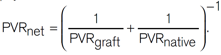
The PVR in the native lung is 14.1 WU and graft lung 2.7 WU. What is the net PVR of the pulmonary circulation?
ANSWER 💡: The correct ansewr is D. PVR of this parallel circuit is calculated by adding the resistance of each lung. It's physics! Here's how it's done. 🤯🤯🤯
Here's the full table of hemodynamics.

The patient underwent successful pulmonary vein stenting and is doing well in follow-up.👍🎉🙂
In summary, this patinet had two different types of pulmonary hypertension after lung transplantation. The native lung had pre-capillary (group 3) and the graft had acquired pulmonary vein stenosis!
The end. Big thanks to our patient for sharing her story. 🙏🙏🙏
ping
@LeaHarperMD
@dr_benoy_n_shah
@ArgaizR
@Nmerke
@Iceman_ex
@RJonesSonoEM
@RaynerHartleyMD
@Thind888
@msiuba
@FH_Verbrugge
@ThinkingCC
@fpmorcerf
@AndrewJSauer
@Wormsy10
@YasMoayedi
@KalagaraHari
@RyanTedfordMD
@LeaHarperMD
@dr_benoy_n_shah
@ArgaizR
@Nmerke
@Iceman_ex
@RJonesSonoEM
@RaynerHartleyMD
@Thind888
@msiuba
@FH_Verbrugge
@ThinkingCC
@fpmorcerf
@AndrewJSauer
@Wormsy10
@YasMoayedi
@KalagaraHari
@RyanTedfordMD
pong
@CalgaryPHdoc
@NephroGuy
@NephroP
@oscaar84
@DrBerticMia
@PulmCrit
@dramcarrillo9
@VatsalTrivediMD
@MSharifpourMD
@katiewiskar
@UAlberta_Sono
@drshahrul80
@EmergencyEcho
@AlbertoAOviedo
@AnishRMitra
@JdBapttiste
@SeguraCardio
@CalgaryPHdoc
@NephroGuy
@NephroP
@oscaar84
@DrBerticMia
@PulmCrit
@dramcarrillo9
@VatsalTrivediMD
@MSharifpourMD
@katiewiskar
@UAlberta_Sono
@drshahrul80
@EmergencyEcho
@AlbertoAOviedo
@AnishRMitra
@JdBapttiste
@SeguraCardio
• • •
Missing some Tweet in this thread? You can try to
force a refresh


