💥‘Pseudohyperkalemia’ Tweetorial
⚡️Pseudohyperkalemia
⚡️Seasonal Pseudohyperkalemia
⚡️Reverse Pseudohyperkalemia
Let’s review these conditions
1/
#Onconephrology
#Hyperkalemia
⚡️Pseudohyperkalemia
⚡️Seasonal Pseudohyperkalemia
⚡️Reverse Pseudohyperkalemia
Let’s review these conditions
1/
#Onconephrology
#Hyperkalemia
⚡️An important point to remember is that 98% of the potassium (K) stores in the body are intracellular so even a small amount of K released from the cells can significantly affect the concentration of ‘measured’ extracellular potassium
2/
2/
⚡️When blood is drawn to measure potassium, you are measuring ‘extracellular’ potassium concentration and NOT intracellular potassium concentration
3/
3/
⚡️Pseudohyperkalemia is when measured potassium concentration in the blood sample is higher than the actual
extracellular potassium in the body because of factors that cause potassium to move out of the cells during or after blood specimen collection
4/
extracellular potassium in the body because of factors that cause potassium to move out of the cells during or after blood specimen collection
4/
⚡️This is why no manifestations of hyperkalemia such as EKG changes are seen in patients with pseudohyperkalemia
⚡️Pseudohyperkalemia is an
in vitro phenomenon
⚡️If Pseudohyperkalemia is not recognized then it can lead to unwarranted therapies
5/
⚡️Pseudohyperkalemia is an
in vitro phenomenon
⚡️If Pseudohyperkalemia is not recognized then it can lead to unwarranted therapies
5/
⚡️What are the factors that lead to Pseudohyperkalemia?
-It can occur due to any factor that leads to potassium leakage out of the cells during or after blood specimen collection
-K can be released from any blood cell: Platelets, WBCs or RBCs
6/
-It can occur due to any factor that leads to potassium leakage out of the cells during or after blood specimen collection
-K can be released from any blood cell: Platelets, WBCs or RBCs
6/
⚡️What causes in vitro cell lysis leading to Pseudohyperkalemia?
-Mechanical Factors
-Temperature
-Chemical Factors
-Time
-Diseases
7/
-Mechanical Factors
-Temperature
-Chemical Factors
-Time
-Diseases
7/
⚡️Pseudohyperkalemia can be caused by cell lysis due to:
-Mechanical factors during or after blood specimen collection such as application of tourniquet, fist clenching, prolonged centrifugation of the blood sample👇🏽
8/

-Mechanical factors during or after blood specimen collection such as application of tourniquet, fist clenching, prolonged centrifugation of the blood sample👇🏽
8/


⚡️Pseudohyperkalemia can occur due to:
-Cold temperature as it inhibits Na-K-ATPase pump leading to leakage of K out of the cell
-Likely why hyperkalemia is seen more often in samples collected in winter season hence the term Seasonal Pseudohyperkalemia👇🏽
9/
-Cold temperature as it inhibits Na-K-ATPase pump leading to leakage of K out of the cell
-Likely why hyperkalemia is seen more often in samples collected in winter season hence the term Seasonal Pseudohyperkalemia👇🏽
9/

⚡️A condition in which low temperature can cause Pseudohyperkalemia👇🏽
-Hereditary stomatocytosis & xerocytosis -> RBC membranes are fragile & sensitive to in vitro K leakage at low temperature👇🏽
-Fresh blood sample at warm temperature will have normal K
10/
-Hereditary stomatocytosis & xerocytosis -> RBC membranes are fragile & sensitive to in vitro K leakage at low temperature👇🏽
-Fresh blood sample at warm temperature will have normal K
10/

⚡️Pseudohyperkalemia can also occur due to:
-Chemical factors: when the antiseptic is not allowed to dry completely before venipuncture then the antiseptic can disrupt cell membrane causing cell lysis & K leakage from the cell👇🏽
11/
-Chemical factors: when the antiseptic is not allowed to dry completely before venipuncture then the antiseptic can disrupt cell membrane causing cell lysis & K leakage from the cell👇🏽
11/

⚡️Pseudohyperkalemia can be a time-dependent phenomenon
-Delayed blood sample processing can exhaust glucose which is needed to generate ATP to maintain the Na-K-ATPase pump which keeps K inside the cell. So Na-K-ATPase malfunction can cause K to leak
12/
-Delayed blood sample processing can exhaust glucose which is needed to generate ATP to maintain the Na-K-ATPase pump which keeps K inside the cell. So Na-K-ATPase malfunction can cause K to leak
12/

⚡️So far all the factors that have been discussed above can lead to Pseudohyperkalemia in both the serum and in the plasma sample
-This is where things get interesting...
13/
-This is where things get interesting...
13/
⚡️Recall the difference between Serum and Plasma?
☄️Serum = Plasma minus the clotting factors
☄️Plasma = Serum plus clotting factors
Plasma is obtained before coagulation of the blood sample & Serum is obtained after coagulation of the blood sample
14/
☄️Serum = Plasma minus the clotting factors
☄️Plasma = Serum plus clotting factors
Plasma is obtained before coagulation of the blood sample & Serum is obtained after coagulation of the blood sample
14/
⚡️Serum sample is collected in a tube with no anticoagulant (such as heparin) so there is blood coagulation whereas Plasma sample is collected in a tube containing an anticoagulant so there is no blood coagulation
‼️ This is an important concept
15/
‼️ This is an important concept
15/
⚡️Now let’s review some diseases that can lead to Pseudohyperkalemia & Reverse Pseudohyperkalemia
-Thrombocytosis or presence of activated platelets
-Leukemia & Lymphoma: an example is Chronic Lymphocytic Leukemia (CLL)
16/
-Thrombocytosis or presence of activated platelets
-Leukemia & Lymphoma: an example is Chronic Lymphocytic Leukemia (CLL)
16/
⚡️Thrombocytosis or presence of activated platelets causes K release from platelets due to platelet degranulation during
in vitro clotting, so K is ⬆️ in serum but NOT in plasma
‼️Serum sample does not have anticoagulant (heparin) but plasma sample does
17/
in vitro clotting, so K is ⬆️ in serum but NOT in plasma
‼️Serum sample does not have anticoagulant (heparin) but plasma sample does
17/

⚡️So Thrombocytosis can lead to falsely high K in serum but NOT in plasma as platelet degranulation during blood clotting is prevented in plasma sample due to presence of an anticoagulant
‼️This is why Pseudohyperkalamia occurs in serum but NOT in plasma
18/
‼️This is why Pseudohyperkalamia occurs in serum but NOT in plasma
18/
⚡️Some investigators have defined Pseudohyperkalemia as a difference of >0.4 mmol/L between serum potassium and plasma potassium
(Potassium being 0.4 mmol/L higher in the serum sample than in the plasma sample)👇🏽
19/
(Potassium being 0.4 mmol/L higher in the serum sample than in the plasma sample)👇🏽
19/

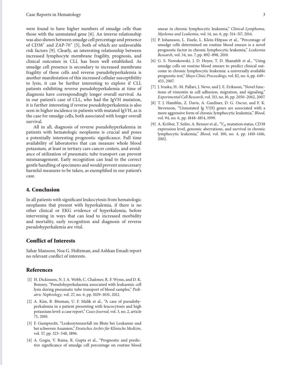
⚡️So what is Reverse Pseudohyperkalemia?
-In conditions like CLL the WBC is very ⬆️, & K can leak from WBCs
-It is thought that the WBC membrane in these conditions is fragile & prone to lysis due to Heparin & during centrifugation👇🏽
20/


-In conditions like CLL the WBC is very ⬆️, & K can leak from WBCs
-It is thought that the WBC membrane in these conditions is fragile & prone to lysis due to Heparin & during centrifugation👇🏽
20/


⚡️Although in CLL you can have falsely high K in both serum & plasma samples but because these WBC membranes are sensitive to heparin you are more likely to see a higher K in Plasma than in the Serum sample hence the term “Reverse Pseudohyperkalemia’
21/
21/
⚡️It is important to be aware of these conditions in order to recognize the difference between serum & plasma potassium concentration and avoid unwarranted therapeutic interventions for pseudohyperkalemia
22/
22/

💥Summary
Pseudohyperkalemia:
⚡️Caused by factors that lead to in vitro cell lysis
⚡️Thrombocytosis can cause Pseudohyperkalemia & CLL can cause Reverse Pseudohyperkalemia
⚡️High K in Pseudohyperkalemia is not real, hence it does not warrant therapy
End/
Pseudohyperkalemia:
⚡️Caused by factors that lead to in vitro cell lysis
⚡️Thrombocytosis can cause Pseudohyperkalemia & CLL can cause Reverse Pseudohyperkalemia
⚡️High K in Pseudohyperkalemia is not real, hence it does not warrant therapy
End/
• • •
Missing some Tweet in this thread? You can try to
force a refresh


