
So; there are often debates regarding ultrasound probe manoeuvres 🤷♂️
Here we go with a graphical Tweetorial, courtesy of myself, @ICUltrasonica and @icmteaching
Hope this helps (you may see these soon in a forthcoming book btw😉
#FOAMed #POCUS #FOAMcc #echofirst 1/7
Here we go with a graphical Tweetorial, courtesy of myself, @ICUltrasonica and @icmteaching
Hope this helps (you may see these soon in a forthcoming book btw😉
#FOAMed #POCUS #FOAMcc #echofirst 1/7
2/7
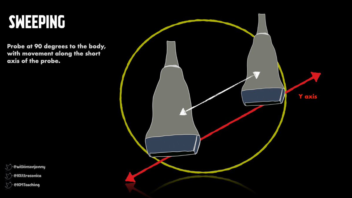
SWEEPING
Here we slide the probe along a slug trail of gel, quite crudely, across a wide area of the body. This is often used to ‘window shop’, for structures we can’t see at first. When they snap into view, we can fine-tune movements 👍 Also allows view of larger organs.
SWEEPING
Here we slide the probe along a slug trail of gel, quite crudely, across a wide area of the body. This is often used to ‘window shop’, for structures we can’t see at first. When they snap into view, we can fine-tune movements 👍 Also allows view of larger organs.
3/7
ROCKING
Classic example here is when we get an apical view of the heart. At first, the heart may not be in line with the scan field. We can ‘swing’ it into view, so it appears more square on the screen. Makes parallel measurements more accurate and things less off-axis🤷♂️
ROCKING
Classic example here is when we get an apical view of the heart. At first, the heart may not be in line with the scan field. We can ‘swing’ it into view, so it appears more square on the screen. Makes parallel measurements more accurate and things less off-axis🤷♂️

4/7
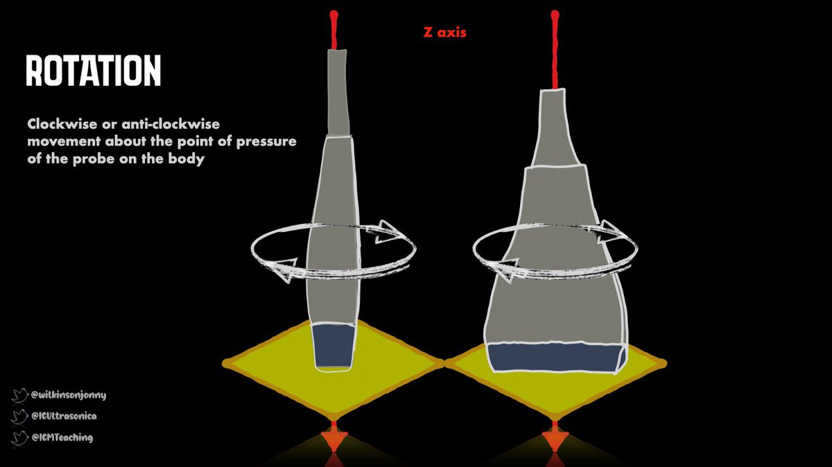
ROTATION
Great for conversion of the view from short axis, to long axis.
Classic example is obtaining a SAX of a blood vessel, then rotation through 90 degrees to view it in LAX for in plane needling during IV access. 🩸 We can do the same viewing nerves for blocks.👏
ROTATION
Great for conversion of the view from short axis, to long axis.
Classic example is obtaining a SAX of a blood vessel, then rotation through 90 degrees to view it in LAX for in plane needling during IV access. 🩸 We can do the same viewing nerves for blocks.👏
5/7
COMPRESSION
E.g. applying gentle downward pressure around the umbilical level to displace bowel gas when we are looking at the abdominal aorta. ⛽️ Another; where we apply pressure when scanning vessels to accentuate the pulsatility of the artery/compress the vein 🌊
COMPRESSION
E.g. applying gentle downward pressure around the umbilical level to displace bowel gas when we are looking at the abdominal aorta. ⛽️ Another; where we apply pressure when scanning vessels to accentuate the pulsatility of the artery/compress the vein 🌊

6/7
SLIDING
Similar to sweeping (this is in short axis), but in the opposite axis (long). We can utilise this when tracing the length of a blood vessel in its long axis for example. This move can also be used to bring off axis images into view.
criticalcarenorthampton.com 👁
SLIDING
Similar to sweeping (this is in short axis), but in the opposite axis (long). We can utilise this when tracing the length of a blood vessel in its long axis for example. This move can also be used to bring off axis images into view.
criticalcarenorthampton.com 👁

7/7
FANNING
The classic here is when viewing a larger organ, like the liver. We essentially guide the beam through the organ by pivoting about a fixed point. It allows better views of subtle architecture that could be missed with broader movements like sweeps 🧹
FANNING
The classic here is when viewing a larger organ, like the liver. We essentially guide the beam through the organ by pivoting about a fixed point. It allows better views of subtle architecture that could be missed with broader movements like sweeps 🧹

Happy Probing to you all and we hope this has been useful🤔🤷♂️🤛👍
Tagging @Manoj_Wickram @zedunow @POCUSAcademy @IMPOCUSFocus @Pocus101 @iceman_ex @ThinkingCC @khaycock2 @load_dependent @PracticalPOCUS @POCUS_Society @NephroP @pocusfoamed @GEHealthcare @ButterflyNetInc
Tagging @Manoj_Wickram @zedunow @POCUSAcademy @IMPOCUSFocus @Pocus101 @iceman_ex @ThinkingCC @khaycock2 @load_dependent @PracticalPOCUS @POCUS_Society @NephroP @pocusfoamed @GEHealthcare @ButterflyNetInc
Here is the overall infographic on ultrasound probe movements for you all to download👍🤛
criticalcarenorthampton.com/pocusgrams/
#FOAMed #POCUS #FOAMcc #echofirst #medtwitter #FOAMus @zedunow @icmteaching @ICUltrasonica
criticalcarenorthampton.com/pocusgrams/
#FOAMed #POCUS #FOAMcc #echofirst #medtwitter #FOAMus @zedunow @icmteaching @ICUltrasonica

• • •
Missing some Tweet in this thread? You can try to
force a refresh


