1/ Thought of doing a quick #tweetorial on image acquisition for #POCUS friends starting to do #VExUS
It's kind of "how I do it" guide and not necessarily optimized for research.
1st: Look at the IVC in both long and short axes
If big, do further scans 👇 #MedEd #FOAMed
It's kind of "how I do it" guide and not necessarily optimized for research.
1st: Look at the IVC in both long and short axes
If big, do further scans 👇 #MedEd #FOAMed

2/ Lateral approach works best to obtain a straight segment of the portal vein (straight = best Doppler shift) and a nice hepatic vein too.
Place transducer approximately in the anterior axillary line pointing towards sternal notch. Then fan antero-posteriorly.
#POCUS #VExUS
Place transducer approximately in the anterior axillary line pointing towards sternal notch. Then fan antero-posteriorly.
#POCUS #VExUS
3/ Forgot what is fanning?
Its also called tilting or some people say, "look" in a particular direction from the same spot.
#POCUS
Its also called tilting or some people say, "look" in a particular direction from the same spot.
#POCUS
4/ When you fan anteriorly, you'll see portal vein and hepatic vein comes into view on posterior fanning. With minor adjustments and a little rotation, both veins can be seen in the same view.
#POCUS
#POCUS
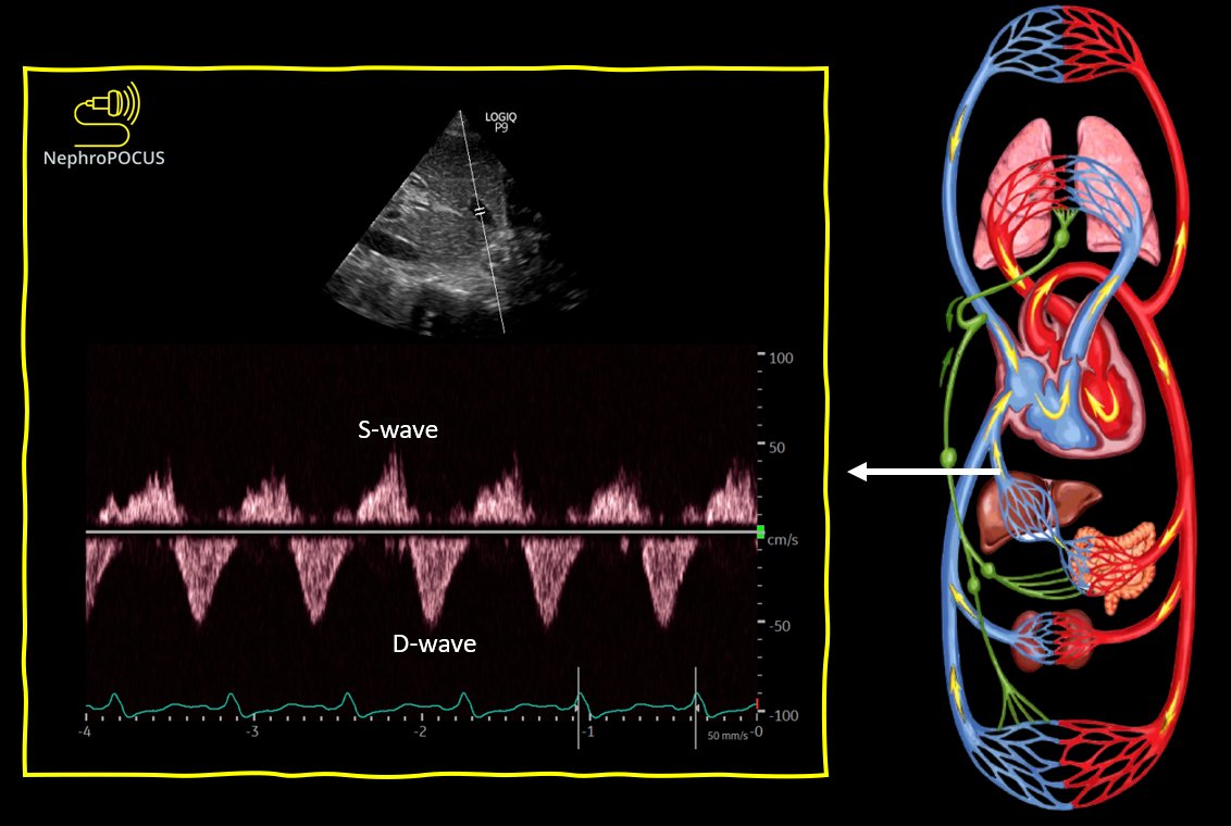
5/ Normal hepatic vein tracing - can appreciate both S and D waves below the baseline👇
@kosmosplatform
@kosmosplatform
6/ These waveforms will be blunted if you ask the patient to hold their breath at end-inspiration 👇
Either do it at end-expiration or during quiet respiration 'when possible'. I know #POCUS is often done in suboptimal conditions & sick patients who cannot follow instructions.
Either do it at end-expiration or during quiet respiration 'when possible'. I know #POCUS is often done in suboptimal conditions & sick patients who cannot follow instructions.
8/ Now lets move on to another window, that is subxiphoid.
In this window, stay slightly towards the right (towards liver); otherwise, bowel gas impedes the views. Remember the abdominal B-lines? #POCUS
In this window, stay slightly towards the right (towards liver); otherwise, bowel gas impedes the views. Remember the abdominal B-lines? #POCUS
9/ In the transverse view, probe orientation marker is to the right (abdomen preset) or to the left (cardiac preset).
If you fan the probe superiorly, hepatic veins-IVC junction comes into view; You'll see portal vein on fanning inferiorly.
#POCUS #VExUS
If you fan the probe superiorly, hepatic veins-IVC junction comes into view; You'll see portal vein on fanning inferiorly.
#POCUS #VExUS
11/ Now long axis view from the subxiphoid window.
If you 'look' to the midline, you'll see aorta. From there, if you fan to the right (towards liver), IVC comes into view and fanning further right will reveal portal vein.
#POCUS #VExUS
If you 'look' to the midline, you'll see aorta. From there, if you fan to the right (towards liver), IVC comes into view and fanning further right will reveal portal vein.
#POCUS #VExUS
12/ Labeled image for better clarity 👇
Though I generally prefer lateral over midline scan, in some patients (obese but lax abdomen; elderly) have great subxiphoid windows.
As mentioned before, hepatic veins often give disturbed flow due to small tributaries here.
Though I generally prefer lateral over midline scan, in some patients (obese but lax abdomen; elderly) have great subxiphoid windows.
As mentioned before, hepatic veins often give disturbed flow due to small tributaries here.

15/ Last component is renal; obviously most of you know how to find kidneys.
Just sweep slightly posterior from the lateral portal vein window, 'look' posteriorly with the probe marker aiming towards posterior axillary line.
Just sweep slightly posterior from the lateral portal vein window, 'look' posteriorly with the probe marker aiming towards posterior axillary line.
16/ As discussed before, use Power Doppler to identify the interlobar vessels when the color pick up is not good.
Here is a normal renal parenchymal tracing (artery on top, vein at the bottom)
#POCUS #VExUS
Here is a normal renal parenchymal tracing (artery on top, vein at the bottom)
#POCUS #VExUS
17/ Hope its helpful. Not as sophisticated as @Wilkinsonjonny 's graphics but a quick curbside teaching 🙃
#POCUS #VExUS
#POCUS #VExUS
• • •
Missing some Tweet in this thread? You can try to
force a refresh


