NephroPOCUS Profile picture
Point-of-care ultrasound #POCUS 📖 | by Abhilash Koratala MD @KoraAbhi, Associate Professor #Nephrology @MCW_Nephrology I👨🏻‍✈️ @POCUSIAPN | X≠ medical advice
Anil Gopinath Profile picture DocPk85 Profile picture Jose De La Flor Profile picture Øystein M. Hansen Profile picture Sarah Owens Profile picture 19 subscribed
Mar 26 14 tweets 4 min read
#eVExUS (= extended #VExUS) includes evaluation of additional veins when the original 3 veins are unobtainable or unreliable for any reason.
Below👇🧵 is an illustrative case showing abnormal pattern in all these veins.
#POCUS #FOAMed #Nephpearls
1⃣ IVC #ultrasound: dilated Image 2⃣ Hepatic vein #Doppler
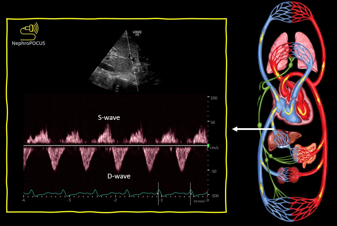
Systolic flow reversal #POCUS #VExUS Image
Nov 15, 2023 5 tweets 2 min read
#echofirst views used to evaluate the right heart.
From ASE comprehensive echocardiography textbook/2010 guidelines
#POCUS #MedEd #CriticalCare #IMPOCUS
(🧵of 5 tweets) Image #POCUS Image
Aug 12, 2023 11 tweets 3 min read
Common #echofirst views used in the #POCUS setting, image acquisition pearls and structures visualized.
Thread below🧵
Courtesy, ASE TTE guidelines 2018
#MedEd #FOAMed #FOAMcc
1⃣ Parasternal window Image 
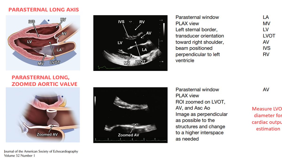
2⃣ Parasternal window continued Image
Jun 27, 2023 21 tweets 6 min read
Focused question: Is HTN in a #dialysis patient always⬆️ intravascular volume? Remove💧?
Clinical: ESRD due to polycystic kidney disease, BP 160s/~100, IVC not well-visualized.
Next step to assess RAP: #VExUS
Below is hepatic vein Doppler
#POCUS #Nephpearls #FOAMcc
Follow the🧵 https://t.co/xZe23bHvJVtwitter.com/i/web/status/1…
2/ 👆What do you think?
May 12, 2023 4 tweets 2 min read
Cardiac #POCUS views - image acquisition pearls from JACC POCUS workbook
#MedTwitter #Nephpearls #FOAMed
Thread🧵below:
1/ Image 2/ Image
May 10, 2023 5 tweets 4 min read
#POCUS enthusiasts, is this IVC normal, abnormal? (asking about possibilities, not definitive conclusions)
#FOAMcc #MedEd #Nephpearls
(Transverse view in thread) IVC transverse #POCUS
Apr 26, 2023 8 tweets 5 min read
Some excellent anatomic illustrations for friends who place central lines.
#POCUS #MedTwitter #Nephpearls #FOAMed
Courtesy: pubmed.ncbi.nlm.nih.gov/27521991/
1. Internal jugular vein (A. Transducer placement in short axis, B. long axis) Image 
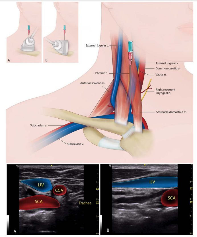
2. The supraclavicular approach to the subclavian vein (A. Transducer in short axis to the vessel, B. long axis)
#POCUS #MedEd Image
Apr 4, 2023 4 tweets 3 min read
#POCUS quiz
1. What tracing did I obtain here?
2. What are some clinical scenarios where doing this would be helpful?
#IMPOCUS #MedEd #Nephpearls Image onlinejase.com/article/S0894-…
#POCUS #MedEd Image
Apr 3, 2023 6 tweets 3 min read
Just plethoric IVC or there is some other notable finding?
#POCUS #MedEd #Nephpearls #POCUS Answer: Pericardial effusion
Cannot comment on tamponade just from this view
Other views in 🧵below. No obvious chamber collapse.
Mar 27, 2023 4 tweets 4 min read
Some helpful illustrations of cardiac support devices for trainees and non-intensivist consultants like me rounding in the ICU.
🔗ccjm.org/content/84/4/2…
#Nephpearls #MedEd #MedTwitter
Also, good to be aware while performing #POCUS ECMO illustrations #MedEd
Feb 1, 2023 5 tweets 4 min read
After yesterday's #POCUS quiz, it's time to reshare these cardiac tamponade infographics.
Courtesy of @ACEP_EUS
🔗acep.org/emultrasound/s…
Set of 3
See 🧵for the rest
#Nephpearls #MedEd #FOAMcc 
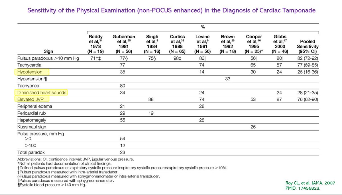
Pulsus paradoxus #echofirst
Feb 1, 2023 4 tweets 3 min read
A 58-year-old woman with no known comorbidities presents with progressive fatigue and shortness of breath x several months. Noted to have bilateral pedal edema; BNP 2,473 pg/mL.
#echofirst 👇❓
Answer and 🔗 to source in thread.
#POCUS #MedEd #FOAMcc Left atrial myxoma -> pulmonary hypertension (RVSP 93 mmHg) -> RV dysfunction (Note obvious RV enlargement ☝️
cvcasejournal.com/article/S2468-…
Jan 30, 2023 7 tweets 5 min read
Another set of cardiac #POCUS #anatomy illustrations. 🧵
#Nephpearls #FOAMed
Source: sciencedirect.com/science/articl…
1⃣ Parasternal long axis 2⃣ Parasternal short axis aortic valve level
#POCUS
Jan 27, 2023 5 tweets 2 min read
#MedEd image of the day. What's wrong with the 🫀?
Clinical: A woman in 40s with h/o severe mitral stenosis secondary to rheumatic heart disease & Afib presented with worsening dyspnea, orthopnea, & chest pain x 7 months
See thread for #echofirst images and source/answer.
#POCUS Image Apical 4-chamber view showing severe right atrial enlargement and annular dilatation of the tricuspid valve during systole.
Dec 28, 2022 8 tweets 6 min read
#POCUS puzzle 🧩of the day. Can you put together the pieces and arrive at an unifying diagnosis? Go through the thread 🧵below.
Context: #dialysis patient, physical examination #FOAMed #Nephrology #MedEd

Image No. 1 👇 Color #POCUS
Dec 6, 2022 4 tweets 3 min read
#POCUS quiz
You are performing physical examination in a patient with suspected fluid overload (plethoric IVC).
Parasternal short axis view demonstrates D-sign. But what's in the RV?
Apical view in thread.
#Nephrology #MedEd #FOAMcc
🔗 to source will be posted in a few hours. Apical #POCUS
Nov 29, 2022 5 tweets 3 min read
#POCUS #tamponade #MedEd
Short axis view and 🔗to source in thread. 👆image shows parasternal long axis view with diastolic collapse of the right ventricle.
👇short axis view at the aortic valve level showing right atrial systolic collapse (arrowhead) and RV diastolic collapse (arrow).
Source: cvcasejournal.com/article/S2468-…
#POCUS #echofirst #MedEd
Nov 18, 2022 10 tweets 5 min read
Basic cardiac #POCUS views image acquisition flash cards.
Courtesy of Philips Healthcare
#MedEd #Nephpearls #echofirst
🧵
1/ #POCUS
Nov 15, 2022 6 tweets 4 min read
Head‑to‑toe #POCUS skills for intensivists in the general and neuro #intensivecareunit population: consensus and expert recommendations of the European Society of Intensive Care Medicine.
🔗pubmed.ncbi.nlm.nih.gov/34787687/
#MedEd #IMPOCUS Summary of the recommendations
#POCUS #Brain
Sep 2, 2022 6 tweets 5 min read
A short 🧵 on hepatic vein #VExUS and key pathologies
From: jacc.org/doi/10.1016/j.…

1/ HV Anatomy & Normal Flow Profile, respiratory variation (forward flow [S,D] ⬆️ during inspiration)

Click ‘ALT’ for normal waveform description

#POCUS #MedEd #Nephrology #IMPOCUS #FOAMed The normal flow profile in ... 2/ A. Tricuspid regurgitation: Systolic flow reversal
B. Tricuspid stenosis: prolonged deceleration time of the D-wave + prominent A-wave

Click ‘ALT’ for further description

#POCUS #VExUS (A) Severe tricuspid regurg...
Aug 22, 2022 22 tweets 18 min read
Time to update the #VExUS resources pinned thread 🧵📌
1/ Tweetorial on image acquisition.
#POCUS #MedEd
2/ The original #VExUS paper
theultrasoundjournal.springeropen.com/articles/10.11… Image