Time for a #POCUS #tweetorial on optimization of Doppler. Very important for #VExUS enthusiasts. #MedEd
1/ Unlike greyscale imaging which depends on amplitude of the returned signal, Doppler depends on frequency information. This graphic explains why perpendicular angle is bad.
1/ Unlike greyscale imaging which depends on amplitude of the returned signal, Doppler depends on frequency information. This graphic explains why perpendicular angle is bad.

2/ other way of saying this, in relevance to color Doppler #POCUS
RBC moving away from the probe = Fr<Ft = negative Doppler shift = Blue color
RBC moving towards = Fr>Ft = positive Doppler shift = Red color
RBC moving away from the probe = Fr<Ft = negative Doppler shift = Blue color
RBC moving towards = Fr>Ft = positive Doppler shift = Red color
Rest of the images/videos from this excellent paper: pubs.rsna.org/doi/10.1148/rg…
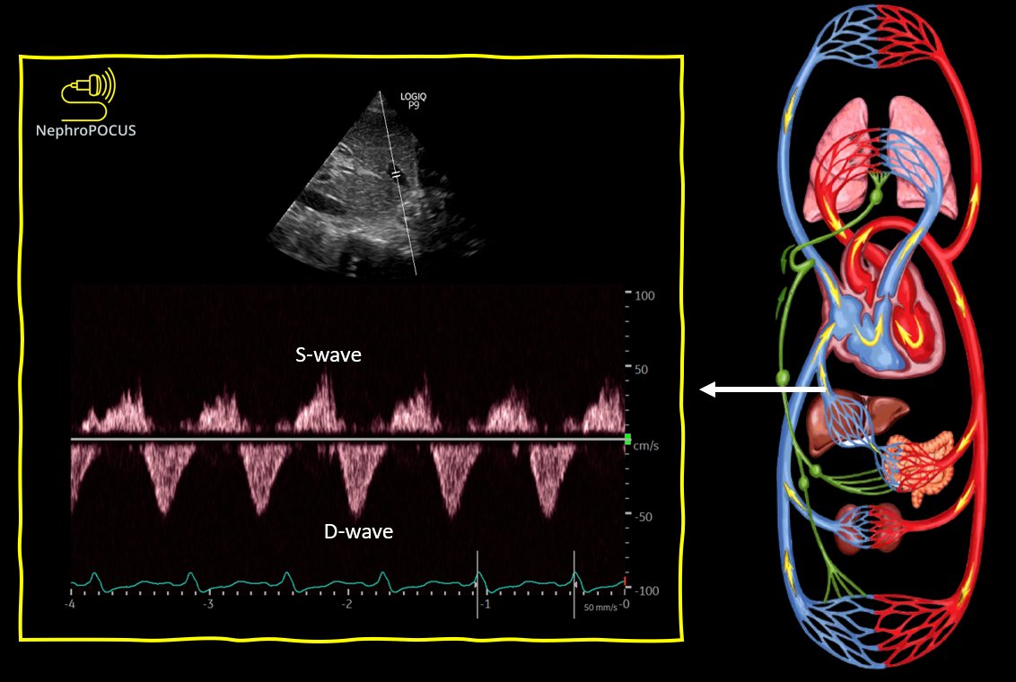
3/ Anatomy (components) of a spectral Doppler waveform (carotid shown)👇
Above baseline is like red on color (towards probe), below = blue. As 0 degree angle is not always possible, <60 is considered OK.
3/ Anatomy (components) of a spectral Doppler waveform (carotid shown)👇
Above baseline is like red on color (towards probe), below = blue. As 0 degree angle is not always possible, <60 is considered OK.

5/ Now coming to optimization:
What if you are not seeing any color? Check maybe the gain is too low to display anything. #POCUS
What if you are not seeing any color? Check maybe the gain is too low to display anything. #POCUS
6/ What happens when the gain is too high? #POCUS
7/ Choosing the appropriate wall filter: wall filters filter out noise due to movement of the vessel wall. If set too high, they filter even the blood flow causing 'gaps' in color flow as well as spectral tracing.
Following is an illustrative image. Read the figure legend too.
Following is an illustrative image. Read the figure legend too.

8/ Wrong wall filter use in spectral Doppler: Loss of slow vascular flow–related information due to a high wall filter setting.
Long axis scan of the proximal abdominal aorta (Ao) shows near-complete loss of diastolic flow at the high wall filter setting (180 Hz) (circled) 👇
Long axis scan of the proximal abdominal aorta (Ao) shows near-complete loss of diastolic flow at the high wall filter setting (180 Hz) (circled) 👇

9/ Another example: portal vein #VExUS 3POCUS obtained at a high wall filter setting (110 Hz) (circled) shows lower-amplitude velocities filtered out, resulting in loss of spectral information immediately above the baseline.

10/ Optimization of color scale: Very important for #vexus
Often people try to scan portal v. in cardiac preset & the scale is high by default - doesn't catch any flow. Similarly, renal flow can be of very low velocity and decreasing the scale helps.
1-minite video (sound on)
Often people try to scan portal v. in cardiac preset & the scale is high by default - doesn't catch any flow. Similarly, renal flow can be of very low velocity and decreasing the scale helps.
1-minite video (sound on)
11/ Importance of optimizing color flow - another example in the context of a mass.
#POCUS
Same principle applies to spectral Doppler tracing. Pay attention to the scale!
#POCUS
Same principle applies to spectral Doppler tracing. Pay attention to the scale!
12/ Circling back to the importance of angle of insonation #POCUS
Absence of flow at one particular point in the portal vein #VExUS - This is due to the angle of insonation of the ultrasound beam being approximately 90° to this part of the vessel - can be confused with thrombus.
Absence of flow at one particular point in the portal vein #VExUS - This is due to the angle of insonation of the ultrasound beam being approximately 90° to this part of the vessel - can be confused with thrombus.

13/ Detection of color flow also depends on the distance of the target organ from the transducer.
Particularly important when doing kidney #VExUS - depends on patient's body habitus & where you are scanning from.
Nice example 👇 #POCUS
Particularly important when doing kidney #VExUS - depends on patient's body habitus & where you are scanning from.
Nice example 👇 #POCUS

14/ on the other hand, color Doppler scan using a different approach (epigastric), aortic flow is readily detectable; the distance to the aorta is 4 cm.
#POCUS
#POCUS

15/ Remember we said velocity scale optimization also applies to spectral Doppler...
here is a nice example 👇 (sound on) #POCUS
here is a nice example 👇 (sound on) #POCUS
16/ Spectral gain:
Like greyscale and color gain (gain = brightness), spectral Doppler gain also can be adjusted & has an impact on how the trace appears.
U must have noticed that I use different colors (brown, pink etc.) for #VExUS traces - different colors need different gain.
Like greyscale and color gain (gain = brightness), spectral Doppler gain also can be adjusted & has an impact on how the trace appears.
U must have noticed that I use different colors (brown, pink etc.) for #VExUS traces - different colors need different gain.
17/ If there are any novice #VExUS #POCUS users, you may want to read my Doppler posts on the Renal fellow network.
Part 1
renalfellow.org/2020/09/24/bas…
Part 1
renalfellow.org/2020/09/24/bas…
That's all for today. Have a nice weekend!
Read the original article I mentioned if you have time.
#POCUS #VExUS
Read the original article I mentioned if you have time.
#POCUS #VExUS
• • •
Missing some Tweet in this thread? You can try to
force a refresh