Want to *see* how a tumour has evolved and grown? And also how different clones acquired characteristic transcriptional and histopathological features?
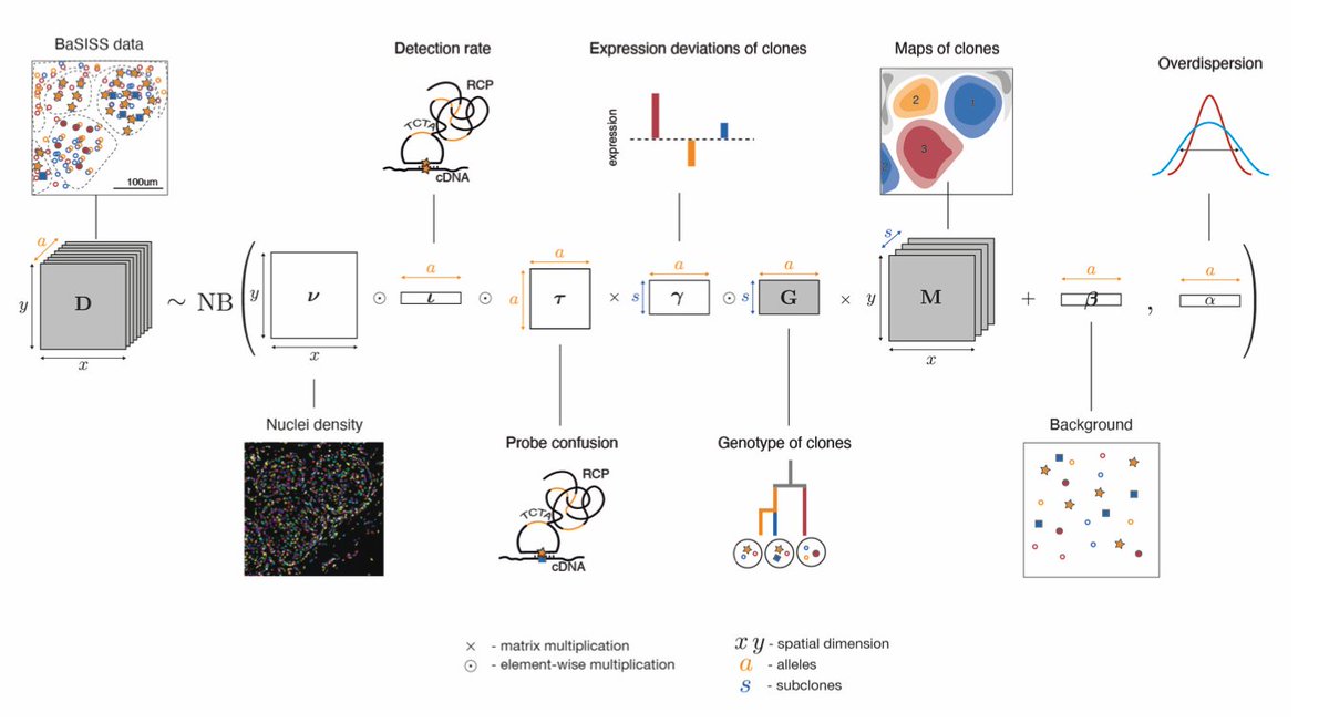
Jessica Svedlund developed a base-specific extension of the in situ sequencing protocol (BaSISS) to detect somatic mutations on a microscopy slide with fluorescently tagged padlock probes. 2/9
These signals are denoised and assembled into microscopic maps of subclonal growth using @LomakinAI's rigorous machine learning model. 3/9
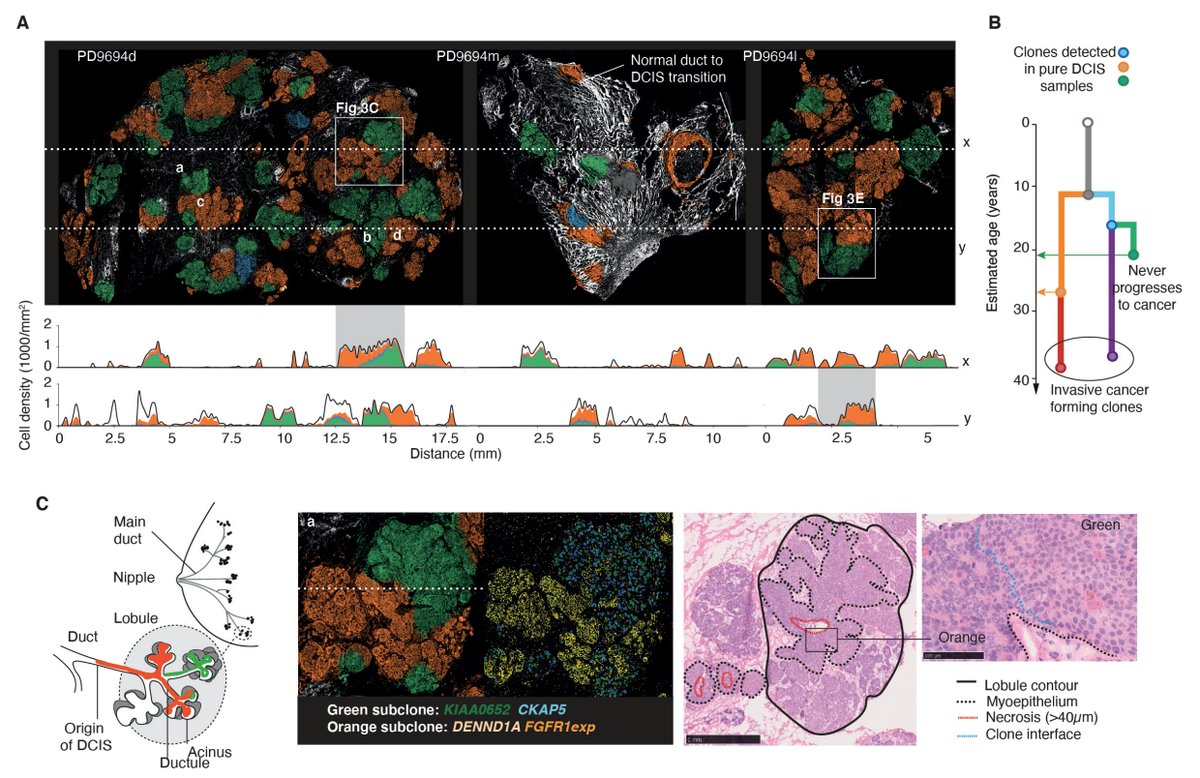
The results are eye watering (for me at least) and show the record of a raging competition of breast cancer clones. The observed clonal growth tightly followed physiological tissue structures. 4/9
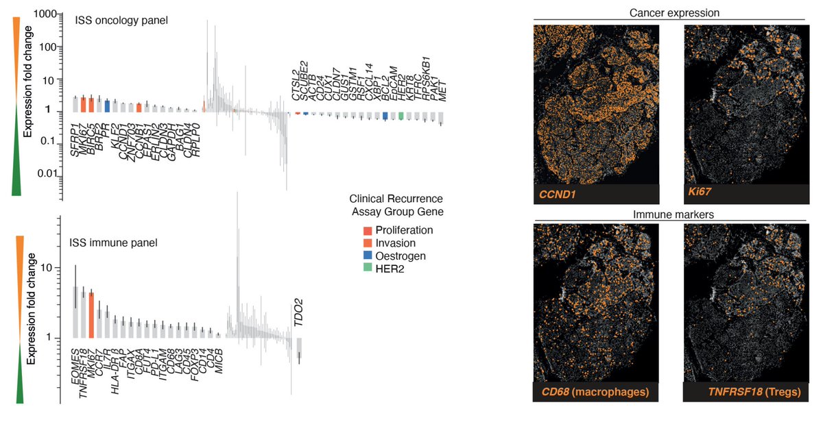
What’s more, layering in ISS probes targeting gene transcripts reveals each clone’s gene expression program and what type of micro environment it surrounds itself with. 5/9
Clones can also have distinct histopathological appearance, further corroborating their phenotypic divergence. 6/9
Mapping these phenotypic patterns onto the underlying phylogeny brings an order into the observed heterogeneity and reveals directional changes as a tumour progresses. 7/9
These snippets highlight the huge potential of spatial genomics and transcriptomics with BaSISS to study cancer evolution. This will enable us to measure, understand - and hopefully one day prevent - the key steps of malignant progression. 8/9
Did the new SARS-CoV-2 B.1.1.7 lineage spread during the English national lockdown? Rising numbers and estimated higher R value suggest so. Together with our colleagues from COG-UK we took a closer look. >> virological.org/t/lineage-spec…
Fitting lineage-agnostic daily PCR test and viral genome data from COG-UK to 382 local authorities we find evidence that B.1.1.7 has spread in a staggering 200/246 of affected LTLAs during the November lockdown (R>1) while at the same time other lineages contracted (R<1). >>
The evidence is therefore overwhelming that B.1.1.7 was repeatedly capable to proliferate under lockdown measures sufficient to suppress other SARS-CoV-2 lineages. B.1.1.7 spread was not an isolated event of general failure of viral containment (both R>1). >>
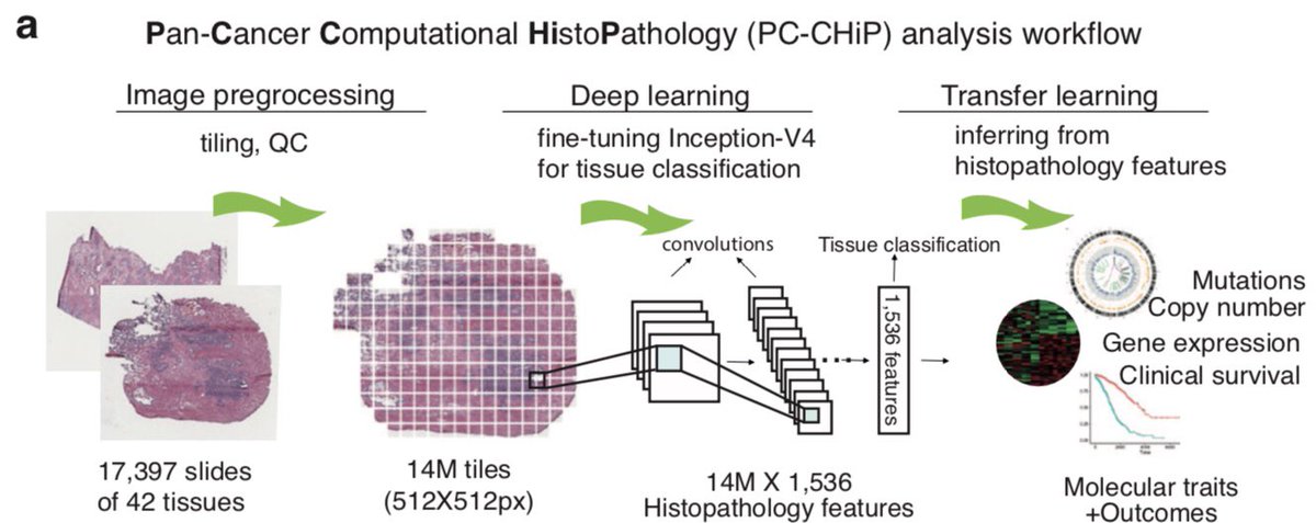
We trained a neural network on 17k tumour slides with known genomics transcriptomics to assess how histopathology, molecular tumour characteristics and survival correspond. 1/8 nature.com/articles/s4301…
This analysis discovered histopathological patterns of 167 different mutations ranging from whole genome duplications to point mutations in cancer driver genes - about 1/4 mutations tested. 2/8
Further, around 40% of the transcriptome is correlated with histopathology reflecting tumour grade and composition. This is probably best illustrated at the example of infiltrating lymphocytes TILs, which can be identified and localised through their expression signature. 3/8
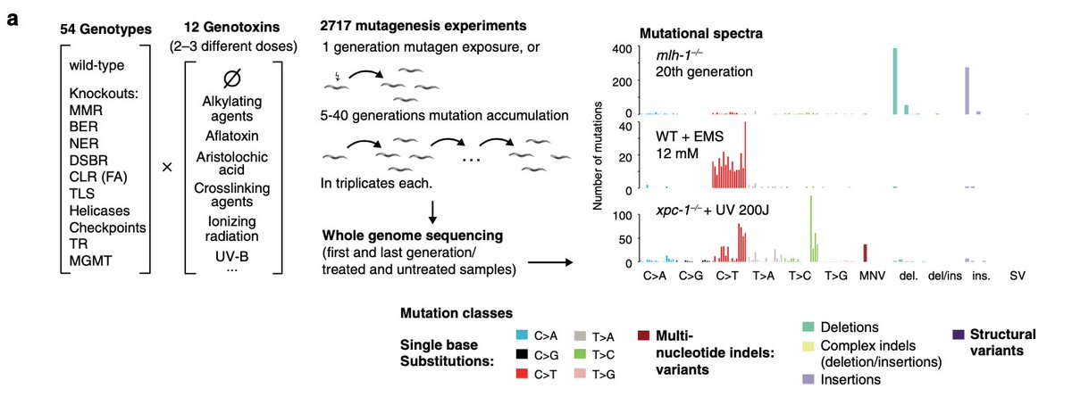
Tired of C19 preprints? Read this: My student @NadezdaVolkova1 & collaborators completely took apart how mutational signatures are sculpted by DNA damage and repair. We grew and sequenced > 2700 worms from 53 repair KO's exposed to 11 mutagens. Phew. 1/7 nature.com/articles/s4146…
Analysing all different combinations, including wildtype and no treatment, allows to map the mutagenic contributions of damage and repair: 9/11 mutagens produce different mutational signatures depending on which repair pathways are acting. This involves 32/53 repair genes. 2/7
We can pin down which elements of a mutational signature are caused by which type of DNA alteration (the same mutagen often produces a variety) – and which repair pathway is involved in mending each type of lesion (usually many pathways operate jointly) 3/7
Hello world. Here’s something interesting: @yufu0413 from my lab trained a deep convolutional neural net in cancer histopathology *and* genomics using 14M images from 17k H&E slides across 28 cancer types. The outcome is stunning. 1/5 biorxiv.org/cgi/content/sh…
The network can predict a good range of genomic alterations, including whole genome duplications. From H&E-images alone. 2/5
It also finds a lot of associations in bulk transcriptome data, deconvolves the signal to find areas on each slide corresponding to molecular cell types such as tumour infiltrating lymphocytes. Entirely automated. 3/5
Here is what we learned recently about somatic evolution and cancer: 1. Mutations arise in virtually every tissue of our body, as a part of normal development and ageing doi.org/10.1101/416339. As a rule of thumb about 1 mutation is introduced at every cell division.
2. Somatic mutations shed light on the first cell divisions in life, informing us about early embryogenesis and how the cells in different parts of our bodies are related to each other via their shared mutations. dx.doi.org/10.1038/nature…dx.doi.org/10.1038/nature…
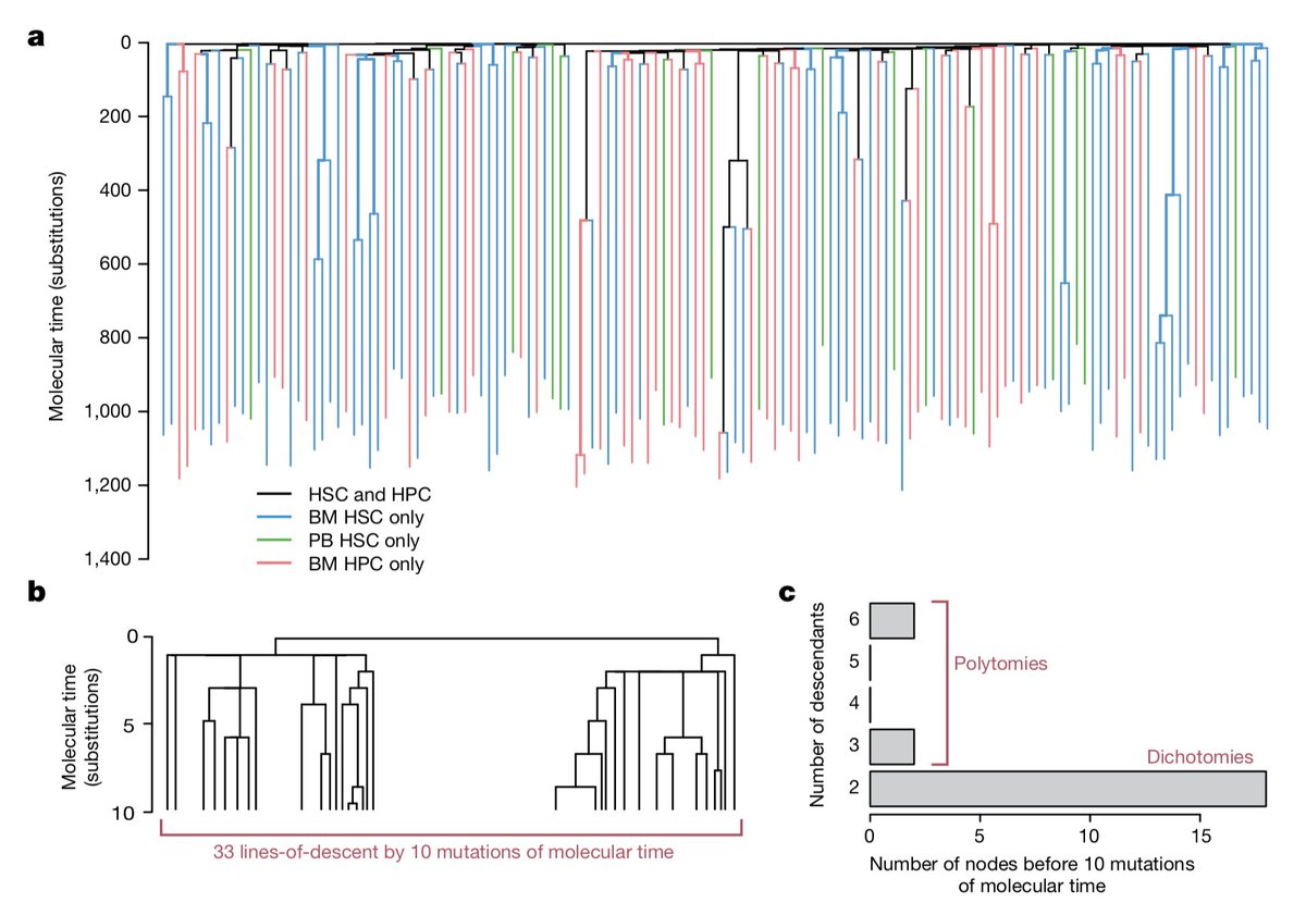
3. In some cases this allows one to draw detailed conclusions about the homeostatic dynamics of an adult organ, exemplified beautifully in recent work on normal blood by Henry Lee-Six, @scienceadvocacy and colleagues. doi.org/10.1038/s41586…
Our work on predicting acute leukaemia based on blood sequencing is out in @nature. Essentially the risk of AML transformation can be determined 5-10yr prior to diagnosis from mutations in blood cells. >> rdcu.be/2Muh
In this study, individuals who progressed to AML harboured more and slightly different pathogenic mutations -- and these affected more cells than in healthy individuals. The time scale is surprising as AML usually manifests rapidly. >>
Yet it is also a demonstration that somatic evolution and neoplastic transformation is common and takes place over long periods. The boundary between benign (ARCH) and malignant (AML) is somewhat blurry, but there is signal to quantify the risk of progression.