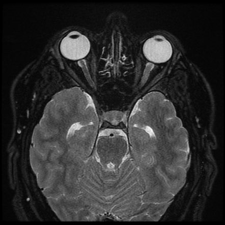
Some optics neuritis pearls in a short #Medtweetorial 🧵…. We all know that optic neuritis is frequently associated with multiple sclerosis (MS). But optic nerve inflammation can exist from autoimmunity, infection, granulomatous disease, paraneoplastic disorders, & demyelination 

Classical ON from MS is unilateral, moderate, painful color vision loss with an afferent pupillary defect & normal fundus examination.
In those with ON, 95% of patients showed unilateral vision loss & 92% had associated retroorbital pain that frequently worsened w/ eye movement.
If there bilateral vision loss, lack of pain, & severe loss of vision, it should raise concern for an alternative inflammatory optic neuropathy
Pay attention to the Vision loss.. Neuromyelitis optica spectrum disorder (NMOSD) & myelin oligodendrocyte glycoprotein (MOG)-IgG optic neuritis cause severe vision loss & are more frequently bilateral.
The absence of an afferent pupillary defect should raise diagnostic concern unless the patient has bilateral involvement or a history of optic neuropathy in the fellow eye. 

In idiopathic ON & ON assoc w/ MS, high-contrast visual acuity loss is moderate, with the majority of patients having acuity better than 20/200. Those w/ neuromyelitis optica spectrum disorder (NMOSD) or MOG-IgG often presents w/ severe vision loss worse than 20/400.
Fundscopic exam for ON is typically normal, with less than 25% of patients presenting w/ disc edema. Significant disc inflammation, disc hemorrhages, or ocular inflammation should raise concern for infection, granulomatous inflammation, or MOG-IgG.
MRI of the orbits is the most sensitive diagnostic test (90%) for optic neuritis; however, a normal orbital MRI scan DOES NOT exclude optic neuritis. Image negative ON. Hmmmmm
What about an ANA? Its not specific for any cause of ON, A pos ANA is more common in patients w/ NMOSD or MOG-IgG ON than in those with MS.
What about the appearance of the On on MRI? Can it clue us in? Yes it can…As Perineural optic nerve enhancement (optic perineuritis) is frequent with MOG-IgG-assoc ON, syphilis, tuberculosis, sarcoidosis, and granulomatosis with polyangiitis (GPA)
And a @k_vaishnani favorite. Check for Bartonella henselae in cases of neuroretinitis in which optic disc edema is accompanied by a macular star of exudates located in a radial pattern around the fovea 

What about the CSF? A mild CSF pleocytosis is frequently observed w/ acute ON; with extensive pleocytosis (>100 cells/mm3) more often in pt w/ MOG-IgG. Pleocytosis of < 50 cells/mm3 is noted in cases of MS-associated ON.
Oligoclonal bands & intrathecal IgG synthesis, hallmarks of optic neuritis associated w/ MS, are uncommon in NMOSD & MOG-IgG-related ON. ncbi.nlm.nih.gov/pmc/articles/P…
• • •
Missing some Tweet in this thread? You can try to
force a refresh


