#IDBoardPearls #IDtwitter #IDPhotoQuiz
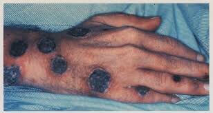
📟 14 YO boy from Ghana with 🤒 & malaise reports a 5 week history of mulriple skin lesions, which are show below. The lesions on his legs are painful & pruritic. He says multiple kids from his school have them too
🧪- VDRL & FTA-ABS +
📟 14 YO boy from Ghana with 🤒 & malaise reports a 5 week history of mulriple skin lesions, which are show below. The lesions on his legs are painful & pruritic. He says multiple kids from his school have them too
🧪- VDRL & FTA-ABS +

❓What organism most likely caused this?
Answer: Treponema pallidum subsp pertenue
🎙Endemic Treponematoes:
🚨Yaws:
🦠- Treponema pallidum subsp pertenue
🗺- Africa, Asia, Latin America, Pacific Islands
Incubation ⏲- 3 wks
Transmission - auto inoculation, close contact with infected lesion
🔎- 🧒🏻👧🏻
Primary Stage -
🎙Endemic Treponematoes:
🚨Yaws:
🦠- Treponema pallidum subsp pertenue
🗺- Africa, Asia, Latin America, Pacific Islands
Incubation ⏲- 3 wks
Transmission - auto inoculation, close contact with infected lesion
🔎- 🧒🏻👧🏻
Primary Stage -
😣, pruritic ulcer that looks like raspberries. These are highly contagious. 🩹 in several mo.
Secondary Stage - appear as primary lesion is healing. Wks- mo. Associated with 🤒 & malaise.
1️⃣Daughter Yaws: lesions are raised
Secondary Stage - appear as primary lesion is healing. Wks- mo. Associated with 🤒 & malaise.
1️⃣Daughter Yaws: lesions are raised

2️⃣🦀 Yaws: plantar or palmer hyperkeratosis causing a painful 🦀like gait

3️⃣ Condyloma lata
4️⃣ Periostitis, osteitis, & dactylitis
Tertiary Stage: destructive bone lesions. Rhinopharyngitits mutilans, hypertrophic periostitis, Saber shins
4️⃣ Periostitis, osteitis, & dactylitis
Tertiary Stage: destructive bone lesions. Rhinopharyngitits mutilans, hypertrophic periostitis, Saber shins

🚨Endemic Syphilis:
🦠- T pallidum subsp endemicum
🗺- North Africa, Arabian Peninsula
Incubation ⏲- 3 wks
Transmission - direct contact with fomites
🔎- 👧🏻🧒🏻
Primary Stage - painless ulcer in oropharynx. Lasts mo-yrs
Secondary Stage - within 3-6 mo.
Mucosal lesions of the
🦠- T pallidum subsp endemicum
🗺- North Africa, Arabian Peninsula
Incubation ⏲- 3 wks
Transmission - direct contact with fomites
🔎- 👧🏻🧒🏻
Primary Stage - painless ulcer in oropharynx. Lasts mo-yrs
Secondary Stage - within 3-6 mo.
Mucosal lesions of the
oropharynx, laryngitis, condyloma lata, angular stomatitis, periostitis
Tertiary Stage: early adulthood
Similar to Yaws
Tertiary Stage: early adulthood
Similar to Yaws

🚨Pinta:
🦠- T pallidum subsp carateum
🗺- Central & South America, Caribbean
Incubation ⏲- 3 wks
Transmission - direct contact with skin lesions
🔎- at any age
Primary Stage - small erythematous papules that coalesce & become hyperpigmented over mo.
Secondary Stage - pintids
🦠- T pallidum subsp carateum
🗺- Central & South America, Caribbean
Incubation ⏲- 3 wks
Transmission - direct contact with skin lesions
🔎- at any age
Primary Stage - small erythematous papules that coalesce & become hyperpigmented over mo.
Secondary Stage - pintids

scaly papules that get darker
Tertiary Stage - lesions become depigmented
Tertiary Stage - lesions become depigmented

🔬- dark field microscopy or immunofluroscence
🧪- nontreponemal (RPR and VDRL) and treponemal (FTA-ABS and TPPPA serology
👇🏼
Won’t be able to differentiate between the 3 and Syphilis
💊- IM benzathine pen G
‼️Leishmaniasis is a protozoal infection caused by the sandfly vector.
🧪- nontreponemal (RPR and VDRL) and treponemal (FTA-ABS and TPPPA serology
👇🏼
Won’t be able to differentiate between the 3 and Syphilis
💊- IM benzathine pen G
‼️Leishmaniasis is a protozoal infection caused by the sandfly vector.
It can cause visceral (Old World, Kala-azar) or cutaneous (New World, mucocutaneous, Espundia) disease depending on the species and location
L major cause CL but the ulcer, which develops wks-mo. after inoculation are usually painless with induration borders. Think 🍕
L major cause CL but the ulcer, which develops wks-mo. after inoculation are usually painless with induration borders. Think 🍕

I should mention that CL wouldn’t be VDRL & FTA-ABS + either. 🙏🏼
• • •
Missing some Tweet in this thread? You can try to
force a refresh