New Lancaster lab publication out in @Nature! This was all @Dabrica’s idea and due to her ingenuity and hard work, with help from @ChiaradiaIlaria, @laupellegrini, and Alex Kalinka. Check it out and see our🧵below!

rdcu.be/cFi7f
Male and female brains differ in their total brain volume. They also show differential susceptibility for some neuropsychiatric disorders. We sought to explore the developmental origin of these differences by generating brain #organoids from male and female stem cell lines.
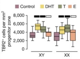
Adding to the cellular complement, we exposed organoids to sex steroids. Addition of androgens (testosterone and DHT) increased the numbers of basal progenitors and their proliferation while estrogen didn’t elicit an observable phenotype.
Viral labelling of radial glia showed that androgens promote symmetric divisions, thus increasing their pool. When DHT was removed from the media, the clone size returned to control levels.
RNA Scope showed presence of androgen receptor in radial glia. Electroporations of constitutively active androgen receptor confirmed increased proliferation capacity of cells. Activating estrogen receptor did not mimic that process.
Bulk and single cell RNA-seq showed involvement of HDACs and mTOR in the generation of androgen-induced phenotype. Drug interference and DHT-mediated rescue confirmed their involvement in androgen signalling. (Both pathways are thought to be involved in ASD)
This happened in the dorsal brain organoids, where excitatory neurons are born. What about ventral, the source of interneurons? Ventral progenitors reacted differently to DHT, without observable increase in intermediate progenitors. Could this contribute to E/I imbalance?
Pulse-chase experiments showed that androgen-increased progenitors resulted in an increase in excitatory neurons. This increase was comparable to the size differences observed between male and female total brain volumes.
Androgens are present in the embryonic brain. At higher levels (in males) our data suggest they specifically increase certain progenitors leading to increased excitatory neurons. This could be one mechanism for the divergence in total brain volumes between males and females.

• • •

Missing some Tweet in this thread? You can try to force a refresh
 

Keep Current with Dr. Madeline Lancaster

Dr. Madeline Lancaster Profile picture

Stay in touch and get notified when new unrolls are available from this author!

Read all threads

This Thread may be Removed Anytime!

PDF

Twitter may remove this content at anytime! Save it as PDF for later use!

Try unrolling a thread yourself!

how to unroll video
  1. Follow @ThreadReaderApp to mention us!

  2. From a Twitter thread mention us with a keyword "unroll"
@threadreaderapp unroll

Practice here first or read more on our help page!

More from @Mad_Lancaster

Oct 27, 2021
I'm thrilled to share our latest published paper in @eLife
where we applied cryo-EM to brain organoids to look at ultrastructure of human axons with unprecedented resolution!

Check out the lovely cryo-CLEM clip below.
And a short 🧵 of what we found.

elifesciences.org/articles/70269
First off, we established a method to culture our air-liquid interface organoid cultures with EM grids to get outgrowth of axon bundles onto the grids. This enables capture of "clean" axons without dendrites like you normally get with cells in vitro.
Then, using correlative light and electron microscopy (CLEM) we could trace axon bundles and focus in on GFP labeled axons within bundles to explore their intracellular architecture.
Read 9 tweets
Mar 24, 2021
📢New Lancaster Lab paper out now! Check it out, we've discovered a cool way evolution has played with cell shape to make our brains BIG! 🧵 cell.com/cell/fulltext/…
This is a question I've been interested in since starting my lab 6 years ago. And so this paper is a really big deal for me and the lab! So where to start...
We know that the human brain is about 3 times bigger than chimps' and gorillas' but why? How?
We can't (nor would we want to) do experiments on developing ape brains, so we approached this question by using brain organoids, little pea-sized replicas of early brain tissue. And when we made organoids from different apes, there was a clear difference in size!
Read 10 tweets
Aug 22, 2020
New preprint from the lab. We’ve joined the fight, and looked at tropism of the virus causing #COVID19 in the brain. Great collaboration with @AnnaAlbecka and Leo James group. Here’s a breakdown of what we find. 🧵

biorxiv.org/content/10.110…
We first look at expression of the viral receptors in human brain organoids and find not much expression, at least at the RNA level, in neural cells. BUT interestingly we find a lot of expression in the choroid plexus. So... what’s the choroid plexus you say?
The choroid plexus (ChP) is what makes your cerebrospinal fluid! It’s also a really important barrier that prevents things from entering the CSF from the blood. So it’s like a gatekeeper, protecting the brain from viruses, toxic compounds and immune cells and factors.
Read 11 tweets

Did Thread Reader help you today?

Support us! We are indie developers!


This site is made by just two indie developers on a laptop doing marketing, support and development! Read more about the story.

Become a Premium Member ($3/month or $30/year) and get exclusive features!

Become Premium

Too expensive? Make a small donation by buying us coffee ($5) or help with server cost ($10)

Donate via Paypal

Or Donate anonymously using crypto!

Ethereum

0xfe58350B80634f60Fa6Dc149a72b4DFbc17D341E copy

Bitcoin

3ATGMxNzCUFzxpMCHL5sWSt4DVtS8UqXpi copy

Thank you for your support!

Follow Us on Twitter!

:(