An in-depth review of metacarpal fractures.
If you're interested in orthopedics you'll definitely want to check this review out!
What is an eponym for this fracture?
If you're interested in orthopedics you'll definitely want to check this review out!
What is an eponym for this fracture?
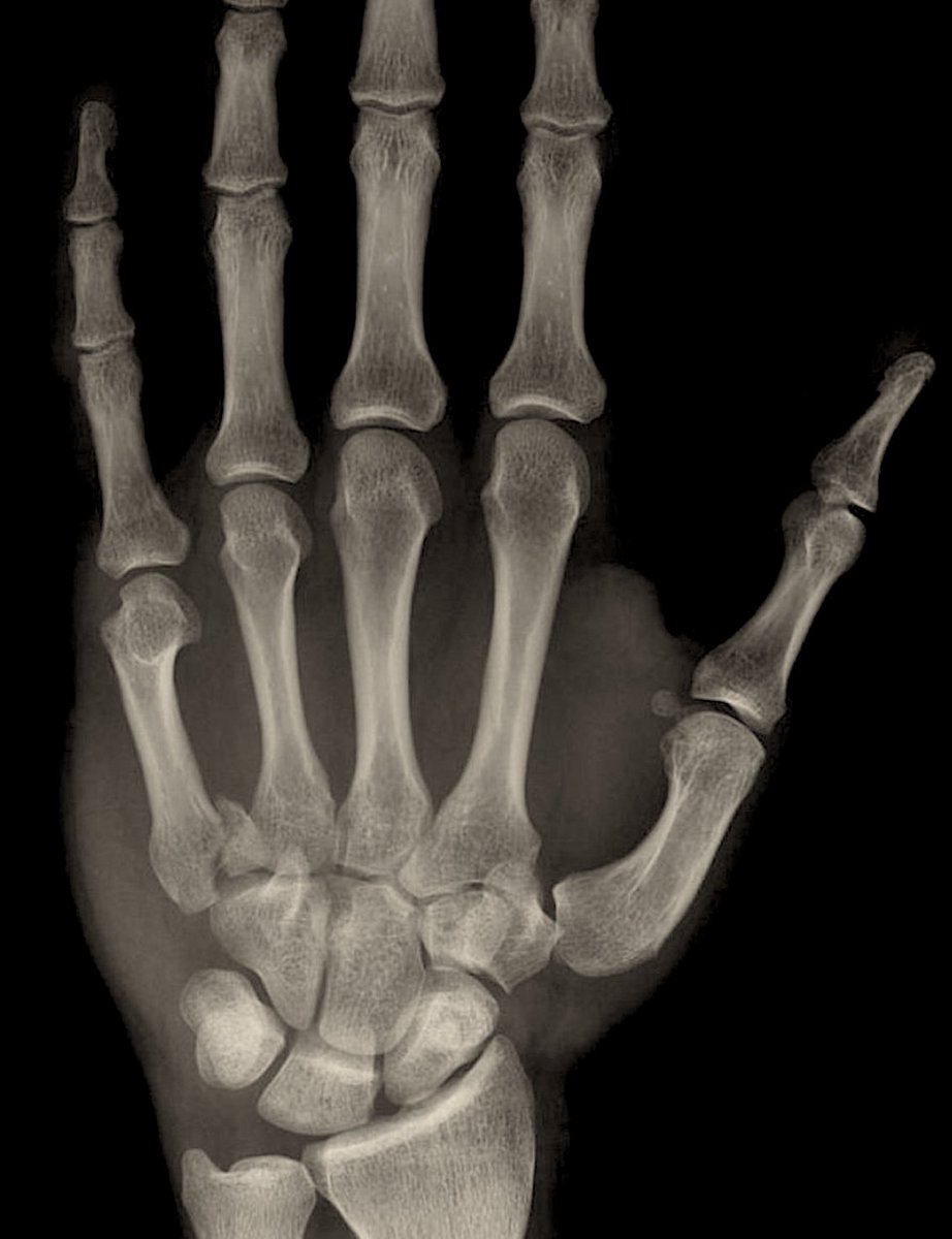
This patient is presenting with an intraarticular fx of the 5th metacarpal base.
This fracture is similar to a Bennett's fx (an intraarticular fx of the 1st metacarpal base).
This fracture goes by a few eponyms: a reverse bennett, baby bennett, or mirrored bennett.
This fracture is similar to a Bennett's fx (an intraarticular fx of the 1st metacarpal base).
This fracture goes by a few eponyms: a reverse bennett, baby bennett, or mirrored bennett.

A Ronaldo fracture is a comminuted fracture of the 1st metacarpal base. (shown above)
Displacement of a Reverse Bennett fracture is due to which of the following muscles?
Displacement of a Reverse Bennett fracture is due to which of the following muscles?
Displacement of a Bennett's fracture is due to the abductor pollicis longus.
Displacement of a Reverse Bennett's fracture is due to the extensor carpi ulnaris.
Displacement of a Reverse Bennett's fracture is due to the extensor carpi ulnaris.

When assessing a pt with a metacarpal fx there are a few things you want to look for:
First, is there an abrasion on the hand, specifically over the MCP. This may indicate a "fight-bite" injury. These injuries are contaminated with oral flora & should be treated with antibiotics
First, is there an abrasion on the hand, specifically over the MCP. This may indicate a "fight-bite" injury. These injuries are contaminated with oral flora & should be treated with antibiotics
Second, you should perform a neurovascular exam to rule out injury to digital nerves/vasculature.
Third, you should assess for deformity. This can be performed by having the patient flex their digits towards the scaphoid tubercle and looking for overlap. (Shown below)
Third, you should assess for deformity. This can be performed by having the patient flex their digits towards the scaphoid tubercle and looking for overlap. (Shown below)

A Boxer's fx occurs when an amateur fighter strikes an object with a flexed wrist resulting in a fracture of the 4th or 5th metacarpal.
If a trained fighter were to fracture a metacarpal it would occur in the 2nd or 3rd metacarpal because they would strike with a neutral wrist.
If a trained fighter were to fracture a metacarpal it would occur in the 2nd or 3rd metacarpal because they would strike with a neutral wrist.

Angulation tolerances for metacarpal shaft fx:
✯ 2nd/3rd: 20°
✯ 4th: 30°
✯ 5th: 40°
Angulation tolerances for metacarpal neck fx:
✯ 2nd/3rd: 15°
✯ 4th: 40°
✯ 5th: 60°
2-5 mm of shortening may be acceptable.
Malrotation is not acceptable
✯ 2nd/3rd: 20°
✯ 4th: 30°
✯ 5th: 40°
Angulation tolerances for metacarpal neck fx:
✯ 2nd/3rd: 15°
✯ 4th: 40°
✯ 5th: 60°
2-5 mm of shortening may be acceptable.
Malrotation is not acceptable

Patients presenting with metacarpal fx may be splinted in an intrinsic plus or "Edinburgh" position:
Wrist slightly extended 15-30°
MCPs are flexed at 80-90°
PIP/DIPs are extended
This is done to help reduce stiffness because the collateral ligaments are shortest in extension.
Wrist slightly extended 15-30°
MCPs are flexed at 80-90°
PIP/DIPs are extended
This is done to help reduce stiffness because the collateral ligaments are shortest in extension.

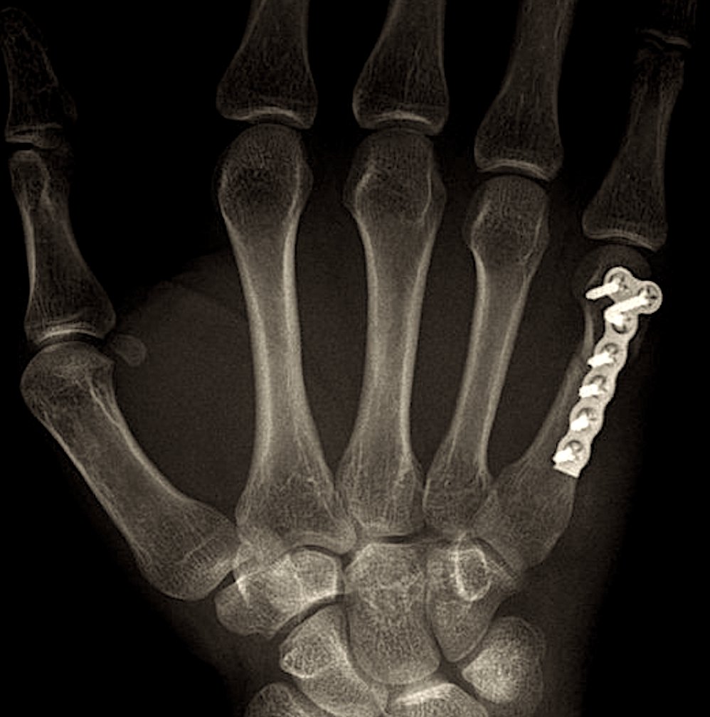
Operative treatment involves closed reduction with percutaneous pinning "CRPP" (left) or ORIF with plate and screws (right).
This depends on the fracture type e.g. comminuted fractures may require ORIF or if reduction cannot be maintained with CRPP.

This depends on the fracture type e.g. comminuted fractures may require ORIF or if reduction cannot be maintained with CRPP.

Complications:
✯ Stiffness: early ROM is important!
✯ Malunion: cosmetic or functional deformity
✯ Infection: look for fight bite injuries
✯ Stiffness: early ROM is important!
✯ Malunion: cosmetic or functional deformity
✯ Infection: look for fight bite injuries
↓
If you enjoyed this review, please like or retweet to help the page grow.
Author: @CSMorford
#MetacarpalFractures #BoxersFracture #Orthopedics #OrthoTwitter #Ortho #Bones #Fractures #Radiology #Tweetorials #MedED #MedicalEducation #MedTwitter #MedStudentTwitter #MedicalStudent
Author: @CSMorford
#MetacarpalFractures #BoxersFracture #Orthopedics #OrthoTwitter #Ortho #Bones #Fractures #Radiology #Tweetorials #MedED #MedicalEducation #MedTwitter #MedStudentTwitter #MedicalStudent
• • •
Missing some Tweet in this thread? You can try to
force a refresh


