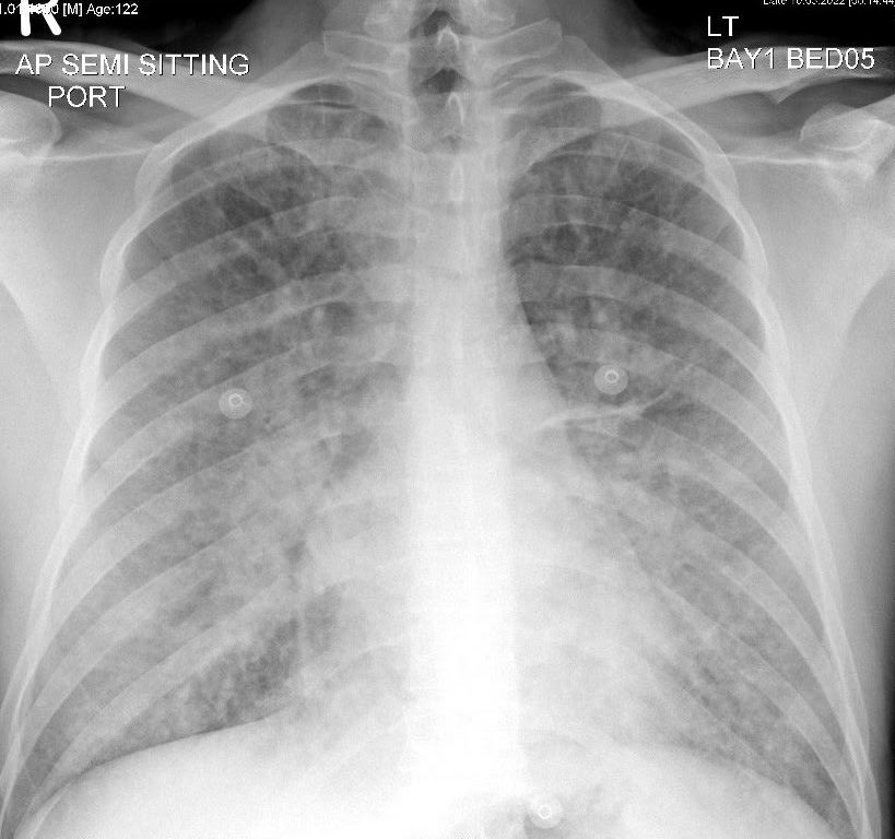
1/n #POCUS case - Pneumothorax. M/20, abrupt left pleuritic chest pain. Absent breath sound left side, hyper-resonant percussion. Absent Pleural sliding on #POCUS . No lung point found, consistent with clinical impression of large pneumothorax.
2/n Normal pleural sliding on #POCUS right anterior thorax
3/n therefore CXR was not a surprise -
4/n Pneumothorax aspirated with 7 Fr CVC catheter, until resistance to aspiration was felt. #POCUS shows return of pleural sliding and #lungpoint
5/n Appearance of lung point on M-mode - Alternating sea-shore and barcode signs
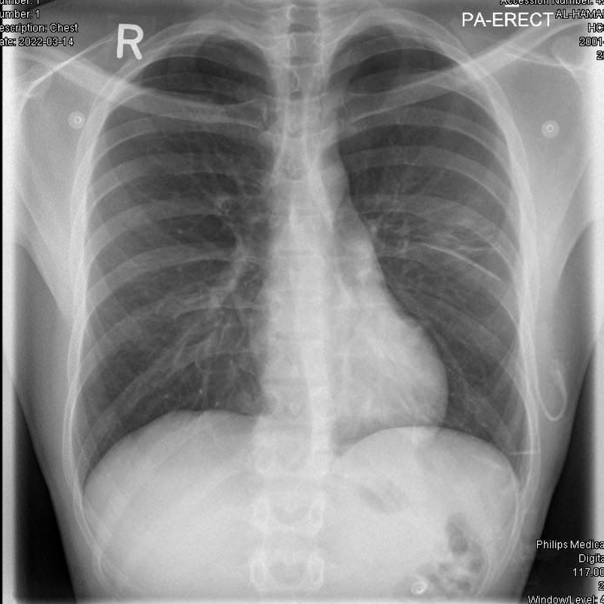
6/6 Observed for 4 hours, repeat #POCUS showed similar findings, CXR repeated, acceptable lung expansion, patient discharged home. ED LOS 5 h.
Thread 1/n #POCUS as a mystery solving tool. M/50, SYNCOPE. Seemed benign orthostatic, and I was upset a Head CT was done. Was expecting it normal, but surprise - reported as left parietal WATERSHED territory infarct. MRI later shows infarct better -
2/n Re-examined carefully, and indeed patient had a homonymous right inferior quadrant-anopia. But WHY does he have watershed infarct unilaterally, from what is a global hypo-perfusion event? Left carotid looks ok...
Thread: 1/3 Clinically anterior glenohumeral dislocation, confirmed by #POCUS within 5 min of arrival
2/3 Pain 10/10 to 0/10 within 5 min of interscalene block
3/3 X-ray was required for followup purposes. Patient walked to x-ray comfortably, and reduced in x-ray suite painlessly and effortlessly. ED LoS 40 min.
Thread, 1/n - Male 75. Acute dyspnea since 1 hour, can't talk, wheezy. SPO2 75%. No previous record. "All that wheezes is not asthma". #POCUS leaves no doubt about pulmonary edema, Dx while patient is being attached to monitor.BP 180/110 mmHg.
2/n CPAP 10 mmHg, IV infusion of nitrate being prepared. Pectus excavatum, parasternal long axis impossible from usual 3/4 ICS. But Apical 4 chamber suggests mild LV systolic dysfunction
3/n As I was about to bolus nitrate 500 mcg - BP drops from 180/110 to 120/70 mmHg. Patient improving with CPAP. So I hold the bolus and try to get more cardiac views. Patient upright plus pectus excavatum, thus not the best quality, but aortic valve appears calcific.
1/n #POCUS is more sensitive than CXR for subtle consolidation detection. M/40, fever, right sided chest pain. Had cough 3 d ago, now resolved.WCC 13k, CRP 230. Normal physical exam.
2/n #POCUS showed bilateral small basal consolidations, B-lines at right base with sub pleural consolidations. Right posterior basal region sub pleural consolidation -