Looks like #POCUS ologists are in a mood to revive old #VExUS posts and tweetorials today.
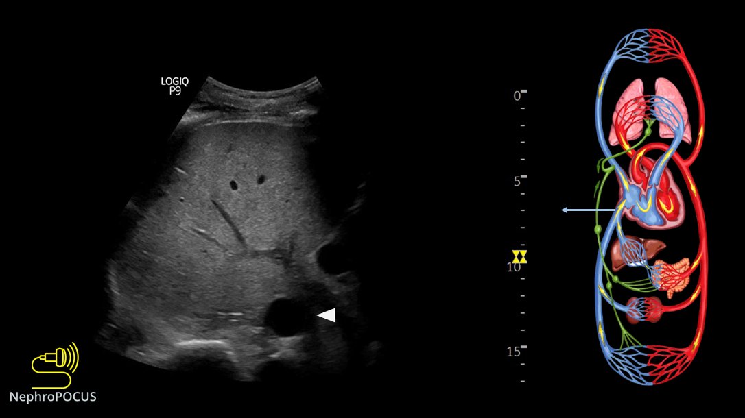
Let me re-share the VExUS flash card(s) 🧵 1. VExUS grading live card #MedEd#IMPOCUS
One of my favorite topics in #POCUS - various effusions seen from different sonographic windows.
Here is a thread 🧵with labeled images for those interested. 1. PLAX view - pericardial effusion - anterior to descending aorta ⚫️below LA), wedge shaped #MedEd#IMPOCUS#Nephpearls
2/ PSAX papillary muscle view
LV is think as many of my patients have #CKD and LVH (as well as mitral annular calcification seen on PLAX view above) #Nephrology#POCUS
3/ Apical 4-chamber view
Sometimes, effusions may be missed due to inadequate visualization of ventricular free wall/lung interference. Always pay attention to the RA area #POCUS
@Rajiv_Sinanan@ThinkingCC 1/ Real reason(s) based on my conversations with people from different backgrounds:
Private practice: "#POCUS sounds interesting but I ain't got time for that. Will consider if I get compensated"
Academia: (not generalizing but most places)
Emphasis is on funded research
@Rajiv_Sinanan@ThinkingCC 2/ Clinical faculty are hired for clinical duties, outreach dialysis etc., which is fair but the problem is educational endeavors are rarely rewarded/provided with protected time. Faculty who want to be #POCUS champions must first learn it (either from institutional experts or
@Rajiv_Sinanan@ThinkingCC 3/ external courses), then integrate into curriculum, take care of administrative stuff/image archiving, prepare didactic material etc., which consume a lot of time and effort. Without dedicated protected time, its almost impossible to continue the initial enthusiasm.
Small thread 🧵illustrating #POCUS based hemodynamic assessment. Relatively a classic case of pulmonary HTN and right heart failure but would like to get some insights from the experts.
2/ Parasternal long axis (PLAX) view demonstrating RV dilatation.
One of the three musketeers is big. Don't know what I'm talking about? Here is a brief reminder: 🔗nephropocus.com/2021/07/12/the…
(Mobile thing in the RVOT is PA catheter; M-mode quiz from this morning is actually this)
3/ Apical 4-chamber view #POCUS
Note how RV is dilated - bigger than LV and forming the cardiac apex.
Inter-atrial septum is bowing to the left indicating high right atrial pressure (not unexpected).
#Nephrology#POCUS short story: 1/ Simple case, want to remind some pitfalls in routine practice.
There was a patient with orthostatic hypotension. Likely neurogenic secondary to amyloidosis.
Read the thread 🧵below: #IMPOCUS#MedEd mini #tweetorial
2/ Physician A orders IV albumin q 6 hours and keeps continuing because: 1. 'Formal' echo from 4 days ago reported a RAP of 3 mmHg = not high 2. 'Documented' weight showed 3 kg drop since admission
Interestingly, documented cumulative fluid balance is +4 L!
3/ Physician B comes in and performs physical examination (#POCUS). Supine BP is relatively low (SBP ~103 mmHg) but nothing different compared to patient-reported figures at home. Pt feels OK, on midodrine.
Here is the IVC👇
1/How can #POCUS#echofirst help in the evaluation of #hyponatremia? #Nephrology consulted for low sodium. Pt received diuretic for shortness of breath (which was possibly due to hiatal hernia as shown on CXR).
2 physicians FELT patient was EUVOLEMIC
Small 🧵below 👇 #MedEd
2/Based on the above labs, it does look like euvolemic hyponatremia (Urine Na is high, Uosm >100 but not too high, BUN not high). BP was 150s systolic.
That's it? Give some salt tablets or UreNa etc.?
No; perform PHYSICAL EXAM (= #POCUS)
IVC was small - doesn't tell much 😬
3/ Next step? Look at the heart, assess stoke volume.
Recent echo LVEF>50%
LV contraction looks good visually
We measured LVOT VTI (couldn't measure LVOT diameter due to chest deformity precluding good PLAX view but its not a problem; VTI is generally enough) #POCUS
Time to discuss some rationale/evidence behind doing #VExUS#POCUS#Nephrology
A short #tweetorial#MedEd 👇 1/ Is fluid overload harmful?
of course yes. Here is a recent meta-analysis.
2/ Does fluid administration affect renal venous flow in asymptomatic but vulnerable patients (#heartfailure)? #POCUS#VExUS
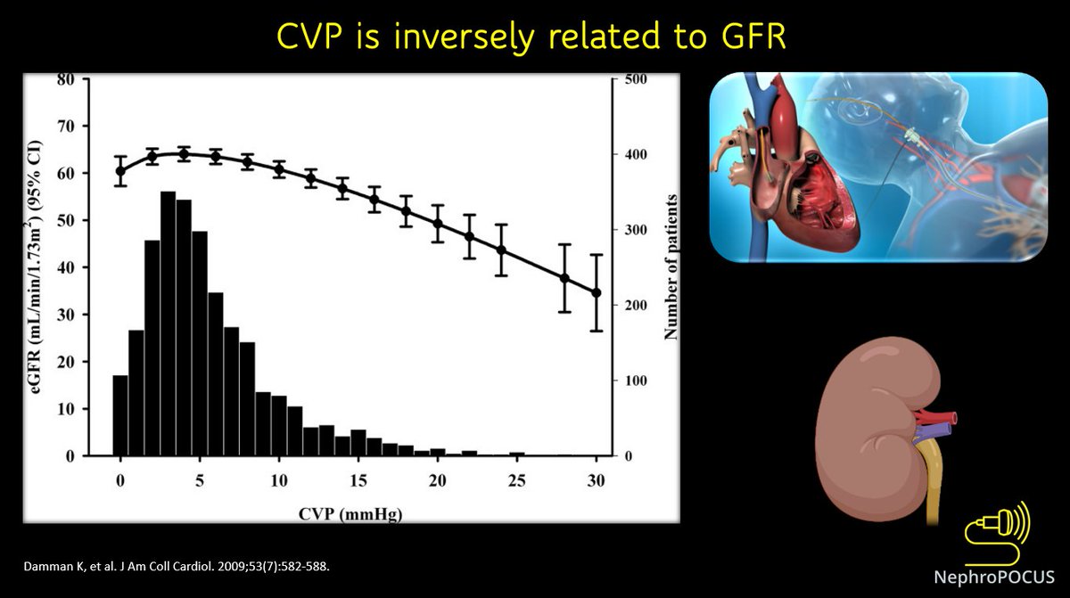
3/ In fact, elevated CVP is associated with reduced GFR.
This 👇is a study in outpatients undergoing right heart cath (N = 2557). In CVP values >6 mm Hg, a steep decrease in GFR was observed.