NephroPOCUS Profile picture
Apr 19 22 tweets 12 min read
One of my favorite topics in #POCUS - various effusions seen from different sonographic windows.
Here is a thread 🧵with labeled images for those interested.
1. PLAX view - pericardial effusion - anterior to descending aorta ⚫️below LA), wedge shaped
#MedEd #IMPOCUS #Nephpearls
2/ PSAX papillary muscle view
LV is think as many of my patients have #CKD and LVH (as well as mitral annular calcification seen on PLAX view above)
#Nephrology #POCUS
3/ Apical 4-chamber view
Sometimes, effusions may be missed due to inadequate visualization of ventricular free wall/lung interference. Always pay attention to the RA area
#POCUS
4/ Subxiphoid view #POCUS
Good view to quickly look for circumferential pericardial effusions
5/ Pericardial effusion is usually detectable on IVC long axis view as well. You just need to pay attention and identify anechoic space between the diaphragm and the heart.
#POCUS
6/ Pleural effusion (left) from PLAX view - posterior to descending aorta. Often the lung is visible floating in the effusion.
#POCUS
7/ Here is another example of left pleural effusion on PLAX view. While lung is not visualized, the effusion is NOT wedge shaped and follows the curvature of the chest wall.
#POCUS
8/ #POCUS = #anatomy + commonsense
Here is the anatomic correlation that explains why descending thoracic aorta is an important landmark to distinguish pericardial and pleural effusions.
9/ Sometimes (thin patients, increased depth) you can see both left and right pleural effusions from the PLAX window. Compare with CT image above.
#POCUS
10/ Pleural effusion from PSAX window. Note the floating lung, which is a big clue in any window.
#POCUS
11/ Another PSAX image showing both pericardial and pleural effusions.
#POCUS
12/ Pleural effusion seen from the apical window.
#POCUS
👆 is called the 'fox tail' sign #POCUS
14/ Apical window demonstrating both pericardial and left pleural effusion.
15/ #POCUS #anatomy correlation for above images
16/ We see a circumferential pericardial effusion in this subxiphoid view. Anything else?
Answer in next tweet
#POCUS
17/ It's ascites
#POCUS #anatomy 👇
18/ Another example of ascites seen in the subxiphoid window. Liver comes into view intermittently, which is a clue to ascites (like lung is to pleural effusion)
#POCUS
19/ The famous Boomerang sign - right pleural effusion seen from the subxiphoid window. This is often confused with ascites.
#POCUS
#POCUS #anatomy correlation for the Boomerang sign 👆
21/ Coming to the end of long 🧵
#POCUS example showing multiple effusions seen from the subxiphoid window.
All the above images can be found in my recent RFN posts. Unfortunately, the gif files are not playing.
🔗renalfellow.org/2021/10/21/ide…
🔗renalfellow.org/2022/04/18/ide…

• • •

Missing some Tweet in this thread? You can try to force a refresh
 

Keep Current with NephroPOCUS

NephroPOCUS Profile picture

Stay in touch and get notified when new unrolls are available from this author!

Read all threads

This Thread may be Removed Anytime!

PDF

Twitter may remove this content at anytime! Save it as PDF for later use!

Try unrolling a thread yourself!

how to unroll video
  1. Follow @ThreadReaderApp to mention us!

  2. From a Twitter thread mention us with a keyword "unroll"
@threadreaderapp unroll

Practice here first or read more on our help page!

More from @NephroP

Apr 18
Looks like #POCUS ologists are in a mood to revive old #VExUS posts and tweetorials today.
Let me re-share the VExUS flash card(s) 🧵
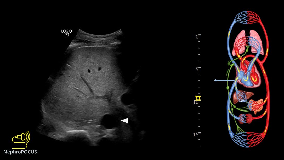
1. VExUS grading live card
#MedEd #IMPOCUS
Hepatic vein #VExUS #POCUS
Portal vein #VExUS #POCUS
Read 5 tweets
Apr 18
@Rajiv_Sinanan @ThinkingCC 1/ Real reason(s) based on my conversations with people from different backgrounds:
Private practice: "#POCUS sounds interesting but I ain't got time for that. Will consider if I get compensated"
Academia: (not generalizing but most places)
Emphasis is on funded research
@Rajiv_Sinanan @ThinkingCC 2/ Clinical faculty are hired for clinical duties, outreach dialysis etc., which is fair but the problem is educational endeavors are rarely rewarded/provided with protected time. Faculty who want to be #POCUS champions must first learn it (either from institutional experts or
@Rajiv_Sinanan @ThinkingCC 3/ external courses), then integrate into curriculum, take care of administrative stuff/image archiving, prepare didactic material etc., which consume a lot of time and effort. Without dedicated protected time, its almost impossible to continue the initial enthusiasm.
Read 8 tweets
Feb 17
Small thread 🧵illustrating #POCUS based hemodynamic assessment. Relatively a classic case of pulmonary HTN and right heart failure but would like to get some insights from the experts.

1/ Parasternal long axis (PSAX) showing D-sign
#VExUS #MedEd #Nephpearls #IMPOCUS
2/ Parasternal long axis (PLAX) view demonstrating RV dilatation.
One of the three musketeers is big. Don't know what I'm talking about? Here is a brief reminder: 🔗nephropocus.com/2021/07/12/the…
(Mobile thing in the RVOT is PA catheter; M-mode quiz from this morning is actually this)
3/ Apical 4-chamber view #POCUS
Note how RV is dilated - bigger than LV and forming the cardiac apex.
Inter-atrial septum is bowing to the left indicating high right atrial pressure (not unexpected).
Read 21 tweets
Jun 29, 2021
#Nephrology #POCUS short story:
1/ Simple case, want to remind some pitfalls in routine practice.
There was a patient with orthostatic hypotension. Likely neurogenic secondary to amyloidosis.
Read the thread 🧵below:
#IMPOCUS #MedEd mini #tweetorial
2/ Physician A orders IV albumin q 6 hours and keeps continuing because:
1. 'Formal' echo from 4 days ago reported a RAP of 3 mmHg = not high
2. 'Documented' weight showed 3 kg drop since admission
Interestingly, documented cumulative fluid balance is +4 L!
3/ Physician B comes in and performs physical examination (#POCUS). Supine BP is relatively low (SBP ~103 mmHg) but nothing different compared to patient-reported figures at home. Pt feels OK, on midodrine.
Here is the IVC👇
Read 9 tweets
May 26, 2021
1/How can #POCUS #echofirst help in the evaluation of #hyponatremia?
#Nephrology consulted for low sodium. Pt received diuretic for shortness of breath (which was possibly due to hiatal hernia as shown on CXR).
2 physicians FELT patient was EUVOLEMIC
Small 🧵below 👇 #MedEd Image
2/Based on the above labs, it does look like euvolemic hyponatremia (Urine Na is high, Uosm >100 but not too high, BUN not high). BP was 150s systolic.
That's it? Give some salt tablets or UreNa etc.?
No; perform PHYSICAL EXAM (= #POCUS)
IVC was small - doesn't tell much 😬
3/ Next step? Look at the heart, assess stoke volume.
Recent echo LVEF>50%
LV contraction looks good visually
We measured LVOT VTI (couldn't measure LVOT diameter due to chest deformity precluding good PLAX view but its not a problem; VTI is generally enough)
#POCUS Image
Read 10 tweets
May 19, 2021
Time to discuss some rationale/evidence behind doing #VExUS #POCUS #Nephrology
A short #tweetorial #MedEd 👇
1/ Is fluid overload harmful?
of course yes. Here is a recent meta-analysis.
2/ Does fluid administration affect renal venous flow in asymptomatic but vulnerable patients (#heartfailure)?
#POCUS #VExUS
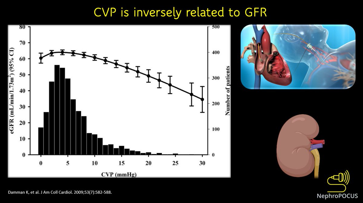
3/ In fact, elevated CVP is associated with reduced GFR.
This 👇is a study in outpatients undergoing right heart cath (N = 2557). In CVP values >6 mm Hg, a steep decrease in GFR was observed.
Read 18 tweets

Did Thread Reader help you today?

Support us! We are indie developers!


This site is made by just two indie developers on a laptop doing marketing, support and development! Read more about the story.

Become a Premium Member ($3/month or $30/year) and get exclusive features!

Become Premium

Don't want to be a Premium member but still want to support us?

Make a small donation by buying us coffee ($5) or help with server cost ($10)

Donate via Paypal

Or Donate anonymously using crypto!

Ethereum

0xfe58350B80634f60Fa6Dc149a72b4DFbc17D341E copy

Bitcoin

3ATGMxNzCUFzxpMCHL5sWSt4DVtS8UqXpi copy

Thank you for your support!

Follow Us on Twitter!

:(