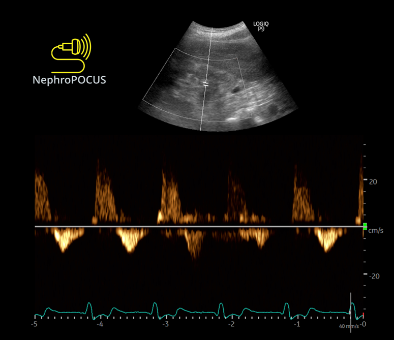
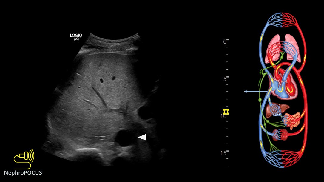
5/ Lungs
Predominantly A-lines, occasional B-lines, but no more than 2 per rib interspace
Representative image👇
6/ So far, venous limb is consistent with congestion/elevated RAP.
Minimally elevated extravascular lung water.
Going to the 🫀 @MDBeni can you please share some pearls for #POCUS users when evaluating prosthetic MV🙏
What you think of this?
7/ 👆Both atria are big; particularly LA with bowing of IAS to the right.
Qualitatively, would you 'suspect' MV stenosis?
👇Gradient across MV. What does this tell?
8/ LV EF looks OK qualitatively.
Let's get LVOT VTI
Looks very high👇
(as you may have noted, there are multiple sample gates; they are automatically appearing when I increase the scale🤦♂️)
9/ As we must take avg. of 3-4 readings, lets see one more. All 4 were similar.
10/ Why VTI is high?🤔
Maybe I should get CWD of LVOT. AS?
👇What does this tell?
11/ Forgot to mention about TR.
Its trace-mild. Couldn't get a good envelope.
#POCUS#echofirst quiz:
In this PLAX view, what structure does the arrow indicate? + what's abnormal about it (if any)?
Clinical: 71y pt with fever/cough, initially thought to be pneumonia, later developed chest pain. BP 134/83 Pulse 106
Will post the answer/source tonight #MedEd
Tweet 1/3
Answer:
Dilated coronary sinus with a mobile echogenicity (vegetation/infective endocarditis in this case)
Full case: 🔗ncbi.nlm.nih.gov/pmc/articles/P…
From #POCUS standpoint, 2 main causes: persistent left superior vena cava & pulmonary HTN. This pt had the former.
2/3 Normally on PLAX view, coronary sinus is barely visible. But when dilated, it can be confused with descending aorta (esp. by users who never encountered this scenario)
One of my favorite topics in #POCUS - various effusions seen from different sonographic windows.
Here is a thread 🧵with labeled images for those interested. 1. PLAX view - pericardial effusion - anterior to descending aorta ⚫️below LA), wedge shaped #MedEd#IMPOCUS#Nephpearls
2/ PSAX papillary muscle view
LV is think as many of my patients have #CKD and LVH (as well as mitral annular calcification seen on PLAX view above) #Nephrology#POCUS
3/ Apical 4-chamber view
Sometimes, effusions may be missed due to inadequate visualization of ventricular free wall/lung interference. Always pay attention to the RA area #POCUS
Looks like #POCUS ologists are in a mood to revive old #VExUS posts and tweetorials today.
Let me re-share the VExUS flash card(s) 🧵 1. VExUS grading live card #MedEd#IMPOCUS
@Rajiv_Sinanan@ThinkingCC 1/ Real reason(s) based on my conversations with people from different backgrounds:
Private practice: "#POCUS sounds interesting but I ain't got time for that. Will consider if I get compensated"
Academia: (not generalizing but most places)
Emphasis is on funded research
@Rajiv_Sinanan@ThinkingCC 2/ Clinical faculty are hired for clinical duties, outreach dialysis etc., which is fair but the problem is educational endeavors are rarely rewarded/provided with protected time. Faculty who want to be #POCUS champions must first learn it (either from institutional experts or
@Rajiv_Sinanan@ThinkingCC 3/ external courses), then integrate into curriculum, take care of administrative stuff/image archiving, prepare didactic material etc., which consume a lot of time and effort. Without dedicated protected time, its almost impossible to continue the initial enthusiasm.
Small thread 🧵illustrating #POCUS based hemodynamic assessment. Relatively a classic case of pulmonary HTN and right heart failure but would like to get some insights from the experts.
2/ Parasternal long axis (PLAX) view demonstrating RV dilatation.
One of the three musketeers is big. Don't know what I'm talking about? Here is a brief reminder: 🔗nephropocus.com/2021/07/12/the…
(Mobile thing in the RVOT is PA catheter; M-mode quiz from this morning is actually this)
3/ Apical 4-chamber view #POCUS
Note how RV is dilated - bigger than LV and forming the cardiac apex.
Inter-atrial septum is bowing to the left indicating high right atrial pressure (not unexpected).
#Nephrology#POCUS short story: 1/ Simple case, want to remind some pitfalls in routine practice.
There was a patient with orthostatic hypotension. Likely neurogenic secondary to amyloidosis.
Read the thread 🧵below: #IMPOCUS#MedEd mini #tweetorial
2/ Physician A orders IV albumin q 6 hours and keeps continuing because: 1. 'Formal' echo from 4 days ago reported a RAP of 3 mmHg = not high 2. 'Documented' weight showed 3 kg drop since admission
Interestingly, documented cumulative fluid balance is +4 L!
3/ Physician B comes in and performs physical examination (#POCUS). Supine BP is relatively low (SBP ~103 mmHg) but nothing different compared to patient-reported figures at home. Pt feels OK, on midodrine.
Here is the IVC👇