A brilliant & timely review by Prof. #DianeEGriffin on the persistence of viral RNA following RNA virus infection - which can be associated with late progressive disease or nonspecific lingering symptoms of post-acute infection syndromes (#PAIS). (1/)
First question addressed is WHERE viral RNA can persist. After a variety of RNA virus infection, viral RNA can persist not only in immune privileged sites (brain, eyes, and testes), but also in blood, lymphoid tissue, joints, respiratory tract, GI tissues, and kidney. (2/)
Consequences of persistent viral RNA may include organ-specific as well as nonspecific postviral syndromes such as long COVID, post-Ebola, and post-polio syndromes, characterized by symptoms including fatigue, headache, muscle pain, and joint pain. (3/)
WHAT is the nature of persistent viral RNA? After all, naked viral RNA would be quickly degraded by host RNases. Persistent RNA may be in the form of; 1) Persistent infectious virus 2) Full-length viral RNA 3) Degraded viral RNA
Evidence for 1) and 2) are discussed. (4/)
Protection of viral RNA may be mediated by ribonucleocapsid (for negative-strand viruses) or association with membranous structures (for positive-strand viruses). More studies are needed to understand HOW vRNA persists. (5/)
Viral RNA persistence reflects the ability of infected cells to avoid elimination by immune mechanisms - innate immunity, antibodies and T cells. This explains viral RNA persistence in long-lived cells like neurons. What about persistence in short-lived cells? (6/)
Short-lived cells like epithelial cells commonly permit rapid cell-to-cell transfer of viral nucleocapsids without release of virus from the cell surface that may foster persistence of viral RNA long after infectious virions can be recovered 🤯 (7/)
Persistent viral RNA can drive chronic innate immune stimulation. If viral RNA is translated, the proteins can also drive chronic adaptive immune stimulation that can lead to prolonged inflammation, as discussed by @jan_choutka@mhornig et al. (8/)
Persistence of viral RNA may also be beneficial if it occurs in the lymph nodes. Measles virus infection in NHP led to vRNA persistence, increase in germinal centers B cells and virus-specific TFH and affinity maturation of antiviral antibody. (9/)
Her review ends with these wise words, “Identification of the role of RNA persistence in late disease could be advanced with longitudinal studies that evaluate treatments that suppress RNA replication and examine their effects on RNA persistence and long-term outcomes.” (10/)
Reflection: persistent viral RNA has been seen post-COVID and after other viral infections. Targeting and getting rid of such RNA may hold key to the treatment of #longCOVID and #MECFS patients who suffer from this pathogenesis. (End)
• • •
Missing some Tweet in this thread? You can try to
force a refresh
With millions of #longCOVID patients, it is becoming better known that even a mild infection can lead to longterm debilitating health problems. SARS-CoV-2 joins the long list of other pathogens that cause post-acute infection syndrome (PAIS). (2/)
What are some common and distinct symptoms associated with PAIS? Strikingly, there are a number of shared symptoms such as excertion intolerance, fatigue, pain, neurological symptoms..etc. Others are more unique to the pathogen that triggered the disease. (3/)
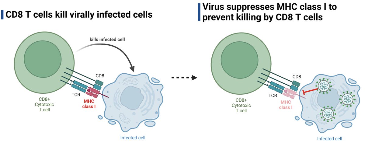
In this study, @MiyuMoriyama et al investigate how well SARS-CoV-2 variants of concern (VOC) suppress MHC I needed for recognition by cytotoxic T cells. This question is important to understand how well the virus limits CD8 killing 🧵(1/) @biorxivpreprint biorxiv.org/content/10.110…
CD8 T cells help fight off viral infection by detecting and killing infected cells. CD8 T cells detect MHC I + viral peptide on infected cells. One of the common tricks viruses use to avoid killing is to inhibit MHC I expression and presentation. (2/)
SARS-CoV-2 is no exception. A previous study showed that SARS-CoV-2 (ancestral) induced MHC I down-regulation in infected cells. They found a key role of ORF8 in this process. (3/)
A Phase 2 clinical trial of oral camostat mesylate during early phase of COVID-19 in outpatients reduced illness course (including fatigue) and prevented loss of smell and taste!
Work of fantastic colleagues at @YaleMed. (1/)
This randomized double-blind placebo-controlled phase 2 trial gave patients (within 3 days of testing PCR+) either oral camostat mesylate or placebo pills, 4x/day for 7 days. Note the lower smell/taste scores (meaning better ability to smell and taste) in camostat group (2/)
Camostat mesylate blocks TMPRSS2, which cleaves the spike protein allowing the virus to fuse with the cell and start to replicate. However, there was no differences in detectable viral RNA levels in patients treated with camostat vs. placebo in nasopharyngeal swab or saliva. (3/)
This new preprint by Stadler et al. integrated data from 37 randomized controlled trials to ask how the timing and dose of passive antibodies (monoclonal Ab & convalescent plasma) predict protection from SARS-CoV-2 disease. A short 🧵 (1/)
Timing: the study found that the earlier the patients were treated with monoclonal antibodies (mAb) or convalescent plasma (CP), the more effective the passive antibodies were in preventing the clinical outcome measured (indicated by right end of line). #TheEarlierTheBetter (2/)
These data are reminiscent of endogenously induced antibody responses against SARS-CoV-2. In patients with fatal COVID, the onset of antiviral antibodies was significantly delayed compared to those who survived COVID. @carolilucas@sneakyvirus1 (3/)
What immune cell features are most predictive of COVID outcomes? @mkuchroo@JcsHuang Patrick Wong et al used ML algorithm Multiscale PHATE to assign each immune cell type in COVID patients a mortality-likelihood score. Latest from @KrishnaswamyLab 💪🏼 (1/) go.nature.com/3K0QCqi
Based on the flow cytometry data on 54 million cells from COVID 168 patients, the low density granulocytes (neutrophils and eosinophils) were the most enriched cell types in patients who had fatal COVID, followed by inflammatory monocytes and certain B cell subsets. (2/)
In contrast, T cells (most of them; see below), NK cells and dendritic cells were associated with the lowest mortality likelihood scores. They are likely protecting the host from lethal disease. (3/)
“COVID toes” are swollen discolored toes (and fingers) that were seen in areas with high incidence of COVID-19, but the cause is unknown. This new study by @JeffGehlhausen et al shows lack of association between covid toes and SARS-CoV-2 infection. 🧵(1/)
We enrolled 23 pandemic chilblains (PC) patients. While there is an association with community COVID cases (blue line) and PC (red bars), only 2 PC patients had evidence of infection by PCR or antibodies. We wondered if people may have missed the time window for testing +ve. (2/)
PCR testing was difficult to access at the time of initial wave (2020). Thus, we employed two distinct measures of antibodies - ELISA and @serimmune SERA assays - against SARS-CoV-2 S, RBD and N. Only 2 of the 23 patients (who were also PCR +ve) had consistent antiviral Abs. (3/)