
🧵 1/9 Thread on constrictive pericarditis. Syncing pathophysio, imaging & hemodynamics to macroscopic findings. @AvrahamCooperMD #CardioTwitter #MedTwitter #echofirst #HeartFailure @YoungDgk @Kardiophil @fuzzymittens @CardioNerds @kardiologie_org @DjawidHashemi @KardiologieHH
https://twitter.com/AvrahamCooperMD/status/1530346310011957248
2/9 Being a fibroelastic sac the pericardium covers & protects the #heart
In constrictive pericarditis:
1️⃣healing of acute pericarditis
2️⃣granulation tissue
3️⃣obliteration of pericardial cavity
4️⃣loss of pericardial elasticity
5️⃣restriction in ventricular filling
#CardioTwitter
In constrictive pericarditis:
1️⃣healing of acute pericarditis
2️⃣granulation tissue
3️⃣obliteration of pericardial cavity
4️⃣loss of pericardial elasticity
5️⃣restriction in ventricular filling
#CardioTwitter
3/9
Correspondingly #cardiacimaging with #cardiacMRI in T1 mapping shows ⬆️ extracellular volume in myocardium, suggestive of global myocardial fibrosis, as shown by doi.org/10.1016%2Fj.jc…
Correspondingly #cardiacimaging with #cardiacMRI in T1 mapping shows ⬆️ extracellular volume in myocardium, suggestive of global myocardial fibrosis, as shown by doi.org/10.1016%2Fj.jc…

4/9
Due to stiff pericardial sac:
1️⃣ intracardiac pressures dissociate from intrathoracic pressure
2️⃣ inspiratory pulm venous pressure decreases, while inspiratory left atrial pressure unchanged
3️⃣ venous return doesn’t change w/ inspiration
☝🏽key difference to cardiac tamponade
Due to stiff pericardial sac:
1️⃣ intracardiac pressures dissociate from intrathoracic pressure
2️⃣ inspiratory pulm venous pressure decreases, while inspiratory left atrial pressure unchanged
3️⃣ venous return doesn’t change w/ inspiration
☝🏽key difference to cardiac tamponade
5/9 As sequelae of this, CP is characterized by specific hemodynamics during inspiration:
1️⃣ LV filling ⬇️
2️⃣ RV filling ⬆️ due to shift of intraventr septum
Echo flow
➡️ insp.: tricuspid valve ⬆️
➡️ exsp.: mitral valve ⬆️ (pictured)
#echofirst #POCUS
doi.org/10.1161/CIRCIM…
1️⃣ LV filling ⬇️
2️⃣ RV filling ⬆️ due to shift of intraventr septum
Echo flow
➡️ insp.: tricuspid valve ⬆️
➡️ exsp.: mitral valve ⬆️ (pictured)
#echofirst #POCUS
doi.org/10.1161/CIRCIM…

6/9 Finally, restriction of diastolic filling results in diastolic pressure equalization in all chambers - a hallmark of CP.
#cardiotwitter
doi.org/10.1016/j.ihj.…
#cardiotwitter
doi.org/10.1016/j.ihj.…

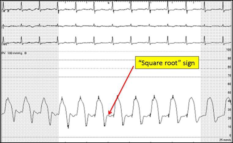
7/9 »Square Root sign« is characteristic in ventr pressure waves.
1️⃣Early ventr diast relaxation isn’t limited
2️⃣Mid-late diast filling is abruptly stopped by CP
3️⃣in late diastole pressures increase until reaching a plateau of equalized pressures.
acc.org/education-and-…
1️⃣Early ventr diast relaxation isn’t limited
2️⃣Mid-late diast filling is abruptly stopped by CP
3️⃣in late diastole pressures increase until reaching a plateau of equalized pressures.
acc.org/education-and-…
8/9 Clinical examination reveals several typical signs
📍kussmaul’s sign - JVP doesn’t decrease during inspiration
📍“pericardial knock“ on auscultation
📍ascites or hepatomegaly due to congestion and elevated filling pressures.
📍kussmaul’s sign - JVP doesn’t decrease during inspiration
📍“pericardial knock“ on auscultation
📍ascites or hepatomegaly due to congestion and elevated filling pressures.
9/9
📍Only definitive management of chronic constrictive pericarditis is pericardiectomy
📍for palliative symptom control or as bridge to surgery diuretics can be used to reduce edema or elevated venous pressures
📍Only definitive management of chronic constrictive pericarditis is pericardiectomy
📍for palliative symptom control or as bridge to surgery diuretics can be used to reduce edema or elevated venous pressures
• • •
Missing some Tweet in this thread? You can try to
force a refresh


