Lea Alhilali, MD Profile picture
Aug 26, 2022 12 tweets 6 min read Read on X
1/”Now your mouth will drop when you see the cord compression we caused,” I said to my fellow looking at our targeted #bloodpatch CT, “But take a deep breath—that’s actually what we want.”
A #tweetorial about CSF leaks & blood patches! #medtwitter #CSFleak #neurotwitter #neurorad Image
2/Epidural blood patches (EBPs) have been around since the 60s. Blood was first injected in the epidural space to try to plug the leak in post-dural puncture HA. It has now been expanded to other CSF leaks. However, controlled studies are lacking & therefore methods vary greatly Image
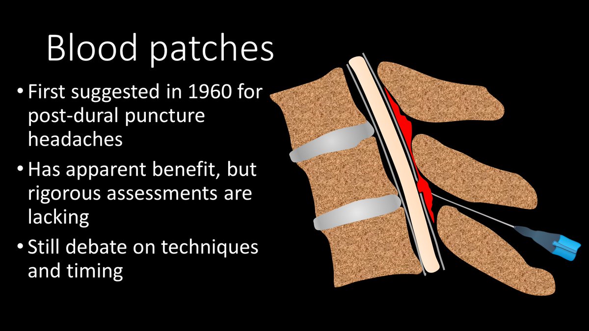
3/No one is sure of how EBPs work. Some believe blood directly plugs the leak site. Other believe it’s a pressure effect--injected blood increases epidural pressure, squeezing the thecal sac like a stress ball, elevating subarachnoid CSF pressure to relieve low pressure HA. Image
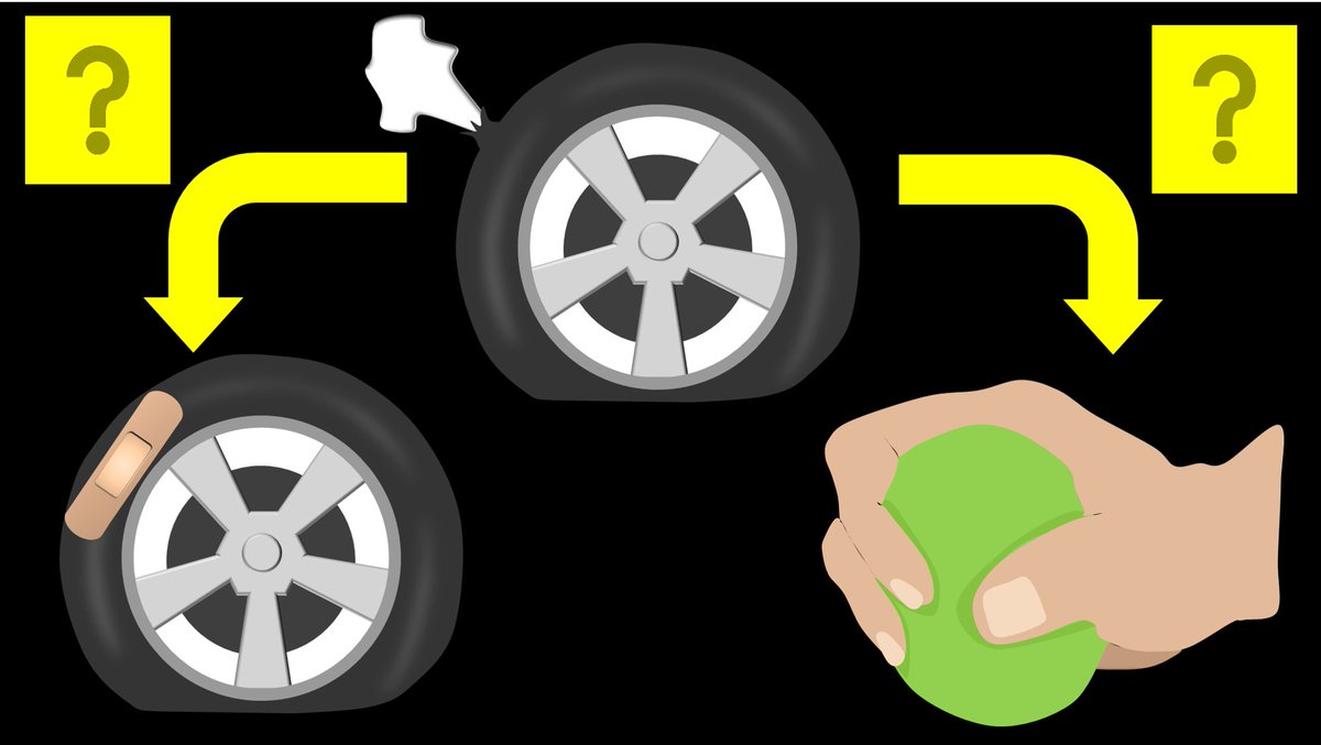
4/In reality, it is probably both mechanisms. The pressure effect is likely what provides the immediate relief from the low pressure HA but the direct plug of the leak is likely what provides the long lasting effectiveness. Image
5/Since direct plugging likely gives long term relief, it’s important to patch the leak site, to increase the likelihood the blood will reach the defect. Finding the leak site could fill a whole other tweetorial. Today we will focus on how to treat the site after it’s found. Image
6/Leaks occur at 3 main sites: (1) Ventrally, usually from an osteophyte spike tearing the dura (2) At the nerve root sleeve, likely related to a leak from a leaking/torn nerve root sleeve diverticulum (3) Dorsally, usually related to a lumbar puncture or spinal intervention Image
7/To get a targeted patch for a ventral leak, a transforaminal approach w/a 22g spinal needle is used to access the ventral epidural space. Care should be taken to avoid the nerve root in the foramen. Both fibrin glue & blood are given to maximize the chance of plugging the leak Image
8/For a leak at the nerve root sleeve, a similar approach for a targeted patch is used, except the needle is stopped short in the foramen and blood/fibrin is given in this region. Image
9/For a nerve root sleeve leak targeted patch, one should see epidural reflux of contrast, to indicate the whole nerve root sleeve has been coated by the patch. For ventral leaks, it is important to confirm that blood has spread across the ventral epidural space to cover the leak Image
10/For a dorsal leak, the traditional interlaminar approach to the epidural space is used. This can be achieved using either fluoroscopy or CT depending on the site.

Choice of injection material/volume can and do vary for all these EBPs depending on the proceduralist Image
11/A significant volume should be given—bc the patch will shrink. I give at least 4cc fibrin & 5-10cc blood—depending on pt tolerance--this guides you. So cord compression is fine, as long as the toes can move. Patch will shrink—like this patch imaged on myelography 3 days later Image
12/Here is a 3D rendering of targeted EBPs/fibrin at 2 levels punctured during spinal stimulator insertion. You can see that over half the canal is filled by the patch. I always tell my fellows a little rhyme: Remember thecal sac compression will lead to symptom regression! Image

• • •

Missing some Tweet in this thread? You can try to force a refresh
 

Keep Current with Lea Alhilali, MD

Lea Alhilali, MD Profile picture

Stay in touch and get notified when new unrolls are available from this author!

Read all threads

This Thread may be Removed Anytime!

PDF

Twitter may remove this content at anytime! Save it as PDF for later use!

Try unrolling a thread yourself!

how to unroll video
  1. Follow @ThreadReaderApp to mention us!

  2. From a Twitter thread mention us with a keyword "unroll"
@threadreaderapp unroll

Practice here first or read more on our help page!

More from @teachplaygrub

May 1
1/Do radiologists sound like they are speaking a different language when they talk about MRI?

T1 shortening what? T2 prolongation who?

Here’s a translation w/an introductory thread to MRI. Image
2/Let’s start w/T1—it is #1 after all! T1 is for anatomy

Since it’s anatomic, brain structures will reflect the same color as real life

So gray matter is gray on T1 & white matter is white on T1

So if you see an image where gray is gray & white is white—you know it’s a T1 Image
3/T1 is also for contrast

Contrast material helps us to see masses

Contrast can’t get into normal brain & spine bc of the blood brain barrier—but masses don’t have a blood brain barrier, so when you give contrast, masses will take it up & light up, making them easier to see. Image
Read 20 tweets
Apr 30
1/Does your ability to remember temporal lobe anatomy seem, well, temporary?

Or are you feeling temporally challenged when it comes to this complex region?

Here’s a thread to help you remember the structures of the temporal lobe! Image
2/Temporal lobe can be divided centrally & peripherally.

Centrally is the hippocampus.

It’s a very old part of the brain & is relatively well preserved going all the way back to rats.

Its main function is memory—getting both rats & us through mazes—including the maze of life Image
3/Peripherally is the neocortex.

Although rats also have neocortex, theirs is much different structurally than humans.

So I like to think of neocortex as providing the newer (neo) functions of the temporal lobes seen in humans: speech, language, visual processing/social cues Image
Read 12 tweets
Mar 25
1/How low can you go??

All the hype nowadays is about high field MRI, but what about low field??

Read on for this month’s @theAJNR SCANtastic for what to know about what may be the next biggest thing in MRI!

ajnr.org/content/47/3/7…Image
2/The growing strength is for larger & larger field strengths for higher & higher resolution

So why would we possible go backwards to lower field strength?

Turns out there are some advantages. Image
3/Low field strength magnets are much for flexible

They can be put in non-traditional settings (clinics) & can also possibly be moved to the bedside

It is truly POC MRI!

But how does it perform? Image
Read 11 tweets
Jan 27
1/The hardest thread yet! Are you up for the challenge?

How stroke perfusion imaging works!

Ever wonder why it’s Tmax & not Tmin?

Here’s what to know from @theAJNR SCANtastic!

ajnr.org/content/47/1/28Image
@TheAJNR 2/Perfusion imaging is based on one principle: When you inject CT or MR intravenous contrast, the contrast flows w/blood & so contrast can be a surrogate marker for blood.

This is key, b/c we can track contrast—it changes CT density or MR signal so we can see where it goes. Image
@TheAJNR 3/So if we can track how contrast gets to the tissue (by changes in CT density or MR signal), then we can approximate how BLOOD is getting to the tissue.

And how much blood is getting to the tissue is what perfusion imaging is all about. Image
Read 19 tweets
Jan 19
1/Do you get a Broca’s aphasia trying remember the location of Broca's area?

Does trying to remember inferior frontal gyrus anatomy leave you speechless?

Don't be at a loss for words when it comes to Broca's area

Here’s a 🧵to help you remember the anatomy of this key region! Image
2/Anatomy of the inferior frontal gyrus (IFG) is best seen on the sagittal images, where it looks like the McDonald’s arches.

So, to find this area on MR, I open the sagittal images & scroll until I see the arches. When it comes to this method of finding the IFG, i’m lovin it. Image
3/Inferior frontal gyrus also looks like a sideways 3, if you prefer. This 3 is helpful bc the inferior frontal gyrus has 3 parts—called pars Image
Read 13 tweets
Dec 5, 2025
1/They say form follows function!

Brain MRI anatomy is best understood in terms of both form & function.

Here’s a short thread to help you to remember important functional brain anatomy--so you truly can clinically correlate! Image
2/Let’s start at the top. At the vertex is the superior frontal gyrus. This is easy to remember, bc it’s at the top—and being at the top is superior. It’s like the superior king at the top of the vertex. Image
3/It is also easy to recognize on imaging. It looks like a big thumb pointing straight up out of the brain. I always look for that thumbs up when I am looking for the superior frontal gyrus (SFG) Image
Read 12 tweets

Did Thread Reader help you today?

Support us! We are indie developers!


This site is made by just two indie developers on a laptop doing marketing, support and development! Read more about the story.

Become a Premium Member ($3/month or $30/year) and get exclusive features!

Become Premium

Don't want to be a Premium member but still want to support us?

Make a small donation by buying us coffee ($5) or help with server cost ($10)

Donate via Paypal

Or Donate anonymously using crypto!

Ethereum

0xfe58350B80634f60Fa6Dc149a72b4DFbc17D341E copy

Bitcoin

3ATGMxNzCUFzxpMCHL5sWSt4DVtS8UqXpi copy

Thank you for your support!

Follow Us!

:(