Nicholas Morris Profile picture
Sep 27 13 tweets 11 min read
1/11 A few years ago, I took care of two young women (some details changed) with similar scans… a brief thread about how MRI can shape a Ddx… but first a poll: Which lesions can restrict diffusion?
2/ Yes… all of these lesions can restrict diffusion. For more info on restricted diffusion see tweets 9-18 in this wonderful tweetorial from @teachplaygrub:
3/So some brief history about the 2 pts. First patient presented to another hospital months ago with R-sided hemiparesis and dysarthria. CSF showed elevated protein but no pleocytosis. Treated several times with steroids with transient improvement but has clinically progressed. Image
4/Second patient developed L hemiparesis over two days. CSF showed neutrophilic pleocytosis with very high protein and normal glucose. Progessed to coma despite high dose steroids (and abx) Image
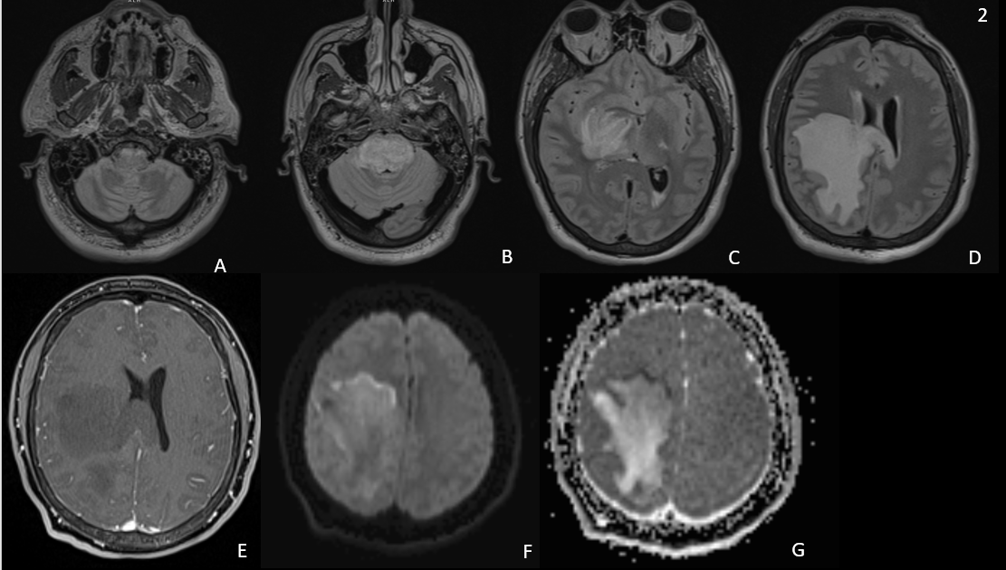
5/ MRIs at first glance very similar. Both with expansile T2 hyperintensity from medulla through basal ganglia. But pt 1’s lesion has gad enhancement (arrows) while pt’s 2 does not. Image
6/Both have areas of restricted diffusion but with very different patterns. Pt 1’s lesion restricts diffusion in the center of the avidly enhancing basal ganglia portion of the lesion. Pt. 2’s restricts diffusion peripherally at the leading edge in the subcortical white matter. ImageImage
7/This is very helpful. Central restricted diffusion is more concerning for highly cellular tumors or abscess. Leading edge restricted diffusion is more concerning for demyelinating lesions (even without enhancement!). n.neurology.org/content/78/21/… Image
8/Pt 2’s biopsy showed an extensive macrophage population with extensive myelin loss with many myelin fragments present within macrophages c/w inflammatory demyelinating lesions, in this case ADEM.
9/ Pt 1’s imaging which we noted could be c/w highly cellular tumor could represent GBM or Primary CNS Lymphoma (PCNSL). PCNSL is more often both supra- and infratentorial and more often had patchy or “stripy” appearance as we see in the cerebellum. rdcu.be/cWrAP Image
10/Pt 1’s biopsy did indeed show Primary CNS Diffuse Large B-cell Lymphoma (DLBCL)
11/11 To summarize, central restricted diffusion in T2 hyperintense lesions more common in highly cellular tumors. Peripheral, or leading edge, restricted diffusion in a T2 hyperintense lesion more common in demyelinating disease. So look carefully!

• • •

Missing some Tweet in this thread? You can try to force a refresh
 

Keep Current with Nicholas Morris

Nicholas Morris Profile picture

Stay in touch and get notified when new unrolls are available from this author!

Read all threads

This Thread may be Removed Anytime!

PDF

Twitter may remove this content at anytime! Save it as PDF for later use!

Try unrolling a thread yourself!

how to unroll video
  1. Follow @ThreadReaderApp to mention us!

  2. From a Twitter thread mention us with a keyword "unroll"
@threadreaderapp unroll

Practice here first or read more on our help page!

More from @namorrismd

May 18, 2021
@UMDNeurosurgery's Dr. Aldrich post hoc analysis shows thick (≥ 4mm) and diffuse (≥ 3 basal cisterns) SAH independently predicts vasospasm-related morbidity and poor 12-wk outcomes. thejns.org/view/journals/…
@claassen_jan used Hijdra grade 3 (completely fills a cistern) to define "thick blood" in Fisher scale revisited paper + showed additive effect of b/l IVH. ahajournals.org/doi/10.1161/hs…
Dr. Jen Frontera, using data from tirilazad RCTs, showed that thick blood and IVH predict symptomatic vasospasm, but did not have explicit definitions for thin/thick and scored IVH as simply present/absent.pubmed.ncbi.nlm.nih.gov/16823296/
Read 5 tweets
Mar 12, 2021
1/ A trainee approached me re: a "hypothetical" case. A patient p/w acute weakness 90 min after onset of R arm/leg without other neuro deficits. The neurology consultant advised IV tPA after CT/CTA head and neck showed no hemorrhage (and also no LVO). The ED attending balked.
2/ Trainee's ?: Can you have a stroke with only motor involvement? Can you have a stroke that causes weakness of the arm and leg, but not the face?... a short tweetorial
3/ C. Miller Fisher described several lacunar stroke syndromes, including pure motor hemiparesis (PMH). He found the most common anatomical locations responsible for PMH (based on autopsy studies) to be the internal capsule and basis pontis. jamanetwork.com/journals/jaman… ImageImage
Read 13 tweets
Feb 11, 2021
Nice study on Thrombocytopenia and Clinical Outcomes in Intracerebral Hemorrhage. Helps further delineate risks for poor outcome after ICH among patients on antiplatelet agents. ahajournals.org/doi/full/10.11…
Over 1/2 of U.S. adults over 45 take antiplatelet agents (APA). ajpmonline.org/article/S0749-…. Studies show disparate results when investigating effect of APA on outcome. doi:10.1161/01.STR.0000231842.32153.74
doi:10.1161/01.STR.0000196991.03618.31
Plt transfusion is common for APA associated ICH, but not show to improve outcomes. PATCH study actually showed worse outcomes in patients who received platelets.
Read 12 tweets
Feb 5, 2021
We recently published our first paper sharing validity evidence for the development of neurological emergency simulations for assessment. Are you familiar with forms of validity evidence? If you are an educator, you should be! A thread… rdcu.be/ceMm3
Steven Downing wrote a fantastic review on validity as it pertains to assessment in medical education. Let’s review the highlights! pubmed.ncbi.nlm.nih.gov/14506816/
As Downing states, validity is the sine qua non of assessment. It is approached as a hypothesis. No assessment is “valid” or “invalid” -> assessments have scores with more or less validity evidence to support interpretations.
Read 17 tweets
Jan 29, 2021
IV glibenclamide shows promise for reducing cerebral edema and appears to be safe. PO glyburide leads to more hypoglycemia, especially if abnl renal fxn. Smaller, more frequent dosing may help. Kudos to @MikeA_42 for pushing this through to publication. sciencedirect.com/science/articl…
@jessestokum performed work in Dr. Marc Simard's lab demonstrating how SUR1-TRPM4 and AQP4 form a complex that amplifies ion/water osmotic coupling and drives astrocyte swelling after brain injury. @UMDNeurosurgery . onlinelibrary.wiley.com/doi/full/10.10…
The role of Sur1-TRPM4 and AQP4 in the milieu that drives cerebral edema was recently summarized nicely by @MDNeurocritcare fellow Melissa Pergakis.tandfonline.com/doi/abs/10.108…
Read 15 tweets

Did Thread Reader help you today?

Support us! We are indie developers!


This site is made by just two indie developers on a laptop doing marketing, support and development! Read more about the story.

Become a Premium Member ($3/month or $30/year) and get exclusive features!

Become Premium

Don't want to be a Premium member but still want to support us?

Make a small donation by buying us coffee ($5) or help with server cost ($10)

Donate via Paypal

Or Donate anonymously using crypto!

Ethereum

0xfe58350B80634f60Fa6Dc149a72b4DFbc17D341E copy

Bitcoin

3ATGMxNzCUFzxpMCHL5sWSt4DVtS8UqXpi copy

Thank you for your support!

Follow Us on Twitter!

:(