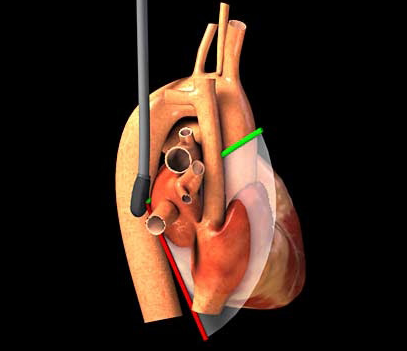
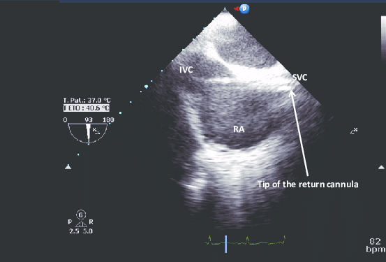
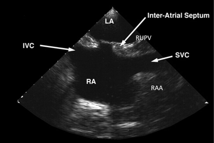
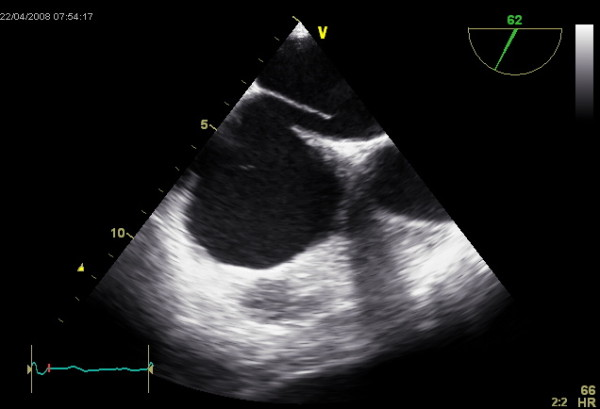
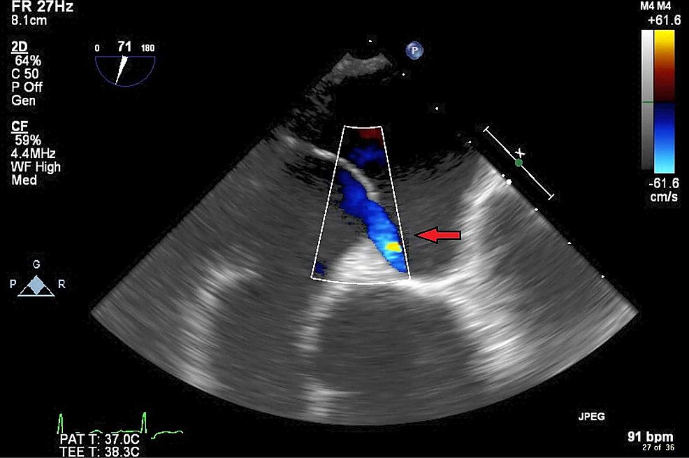
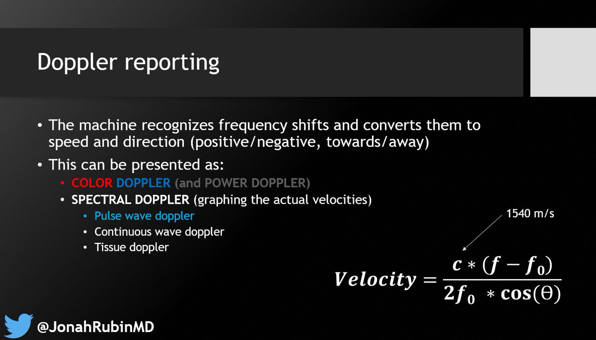
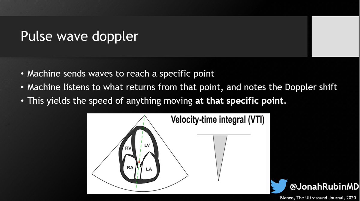
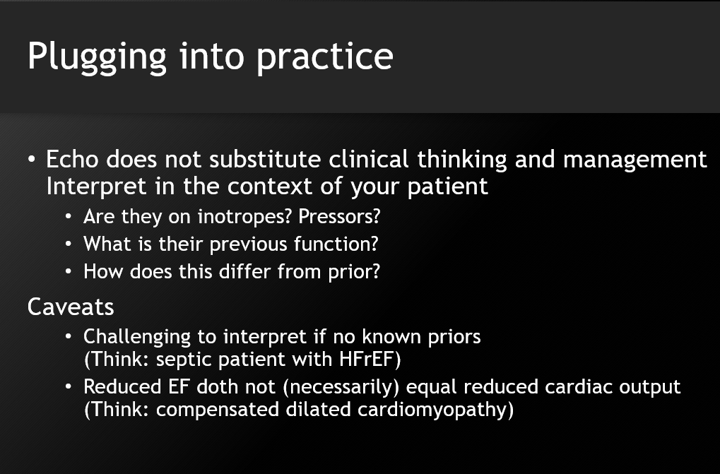
It's a PFO! A few words about this powerful echo window: The bicaval view 🧵 (1/8)

#POCUS #Medtwitter #echofirst #PCCMTwitter #ICUtwitter
This window is attainable only via a transESOPHAGEAL echo (TEE).
Point-of-care TEE is used widely in SICUs worldwide, and MICUs outside the US. You can expect to see them increasingly in US MICUs over the next decade. (2/8)
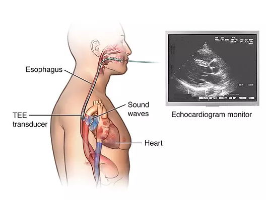
By viewing the heart via the esophagus, windows are often much crisper than when fighting with rib shadows, edematous lungs, and often bandages/wounds (esp. in surgical patients) on the chest. (3/8)
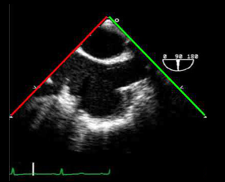
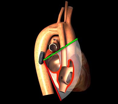
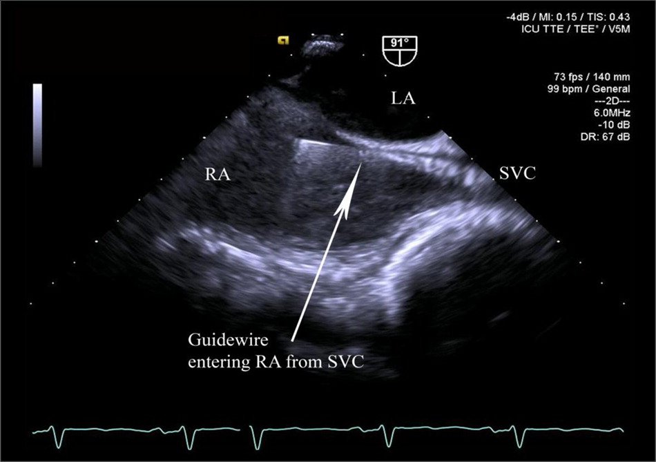
As its name suggests, the bicaval view allows you to see both vena cava - IVC and SVC - in one shot. It's obtained by placing the probe into the mid-esophagus, and rotating the omniplane angle to 90 degrees. Incredible simulator here: pie.med.utoronto.ca/tee/TEE_conten… (4/8)
This view is extremely helpful when placing guidewires/catheters with little room for error - like ECMO cannulas. You can ensure the guidewire is venous, not going into the RV, and for dual lumen ECMO cannulas, confirm placement in both IVC and SVC. (5/8)
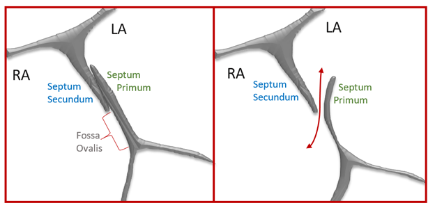
But it can also be seen as a bi*atrial* view because it allows you to see the RA and LA in one shot as well - including, of course, the interatrial septum. Note that the septum has two "beefy" ends, with a thin filament-appearing layer connecting them - the septum primum. (6/8)
Normally, at birth, this adheres to the thicker septum secundum. Sometimes it doesn't truly seal, but the higher pressure of the LA (above the RA) keeps it flush against the septum secundum and closed. But when RA pressure rises above LA pressure, the flap can open ➡️PFO (7/8)
Note - this is not technically an ASD (atrial septal defect) - because the septa are all intact. In an ASD, part of the septum is missing or perforated. Note in this image, and our clip, the flow sneaks underneath the curtain, through the unsealed, patent, foramen ovale! (8/8)
Thanks for reading! As always, thoughts, comments, rebuttals, and additional input always welcome!

• • •

Missing some Tweet in this thread? You can try to force a refresh
 

Keep Current with Jonah Rubin MD

Jonah Rubin MD Profile picture

Stay in touch and get notified when new unrolls are available from this author!

Read all threads

This Thread may be Removed Anytime!

PDF

Twitter may remove this content at anytime! Save it as PDF for later use!

Try unrolling a thread yourself!

how to unroll video
  1. Follow @ThreadReaderApp to mention us!

  2. From a Twitter thread mention us with a keyword "unroll"
@threadreaderapp unroll

Practice here first or read more on our help page!

More from @JonahRubinMD

Oct 6
Cardiac Output by VTI!
Summary slides from our next lecture, broken into 5 steps⤵️
@MGH_PCCM @HarvardPulm @MGHHCICU

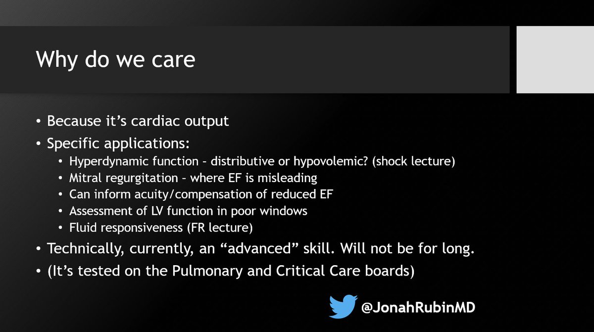
1⃣ Overview & approach - and why you care. CO adds significant detail and nuance to LV function assessment

#POCUS #Medtwitter #PCCMtwitter #echofirst ImageImageImageImage
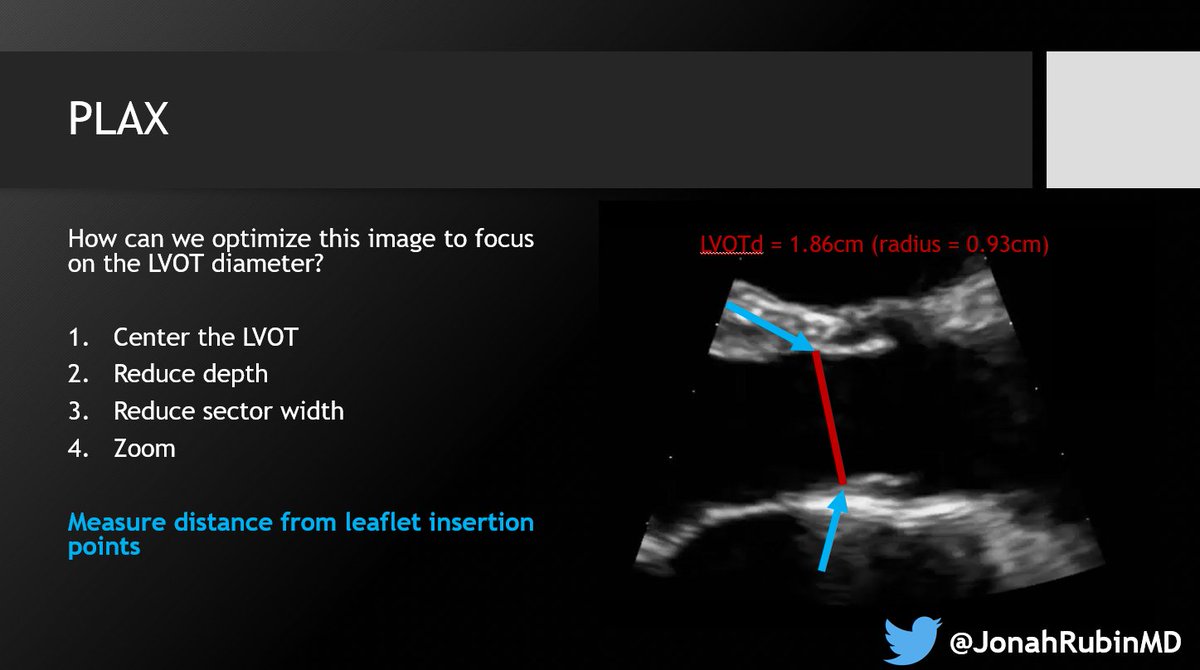
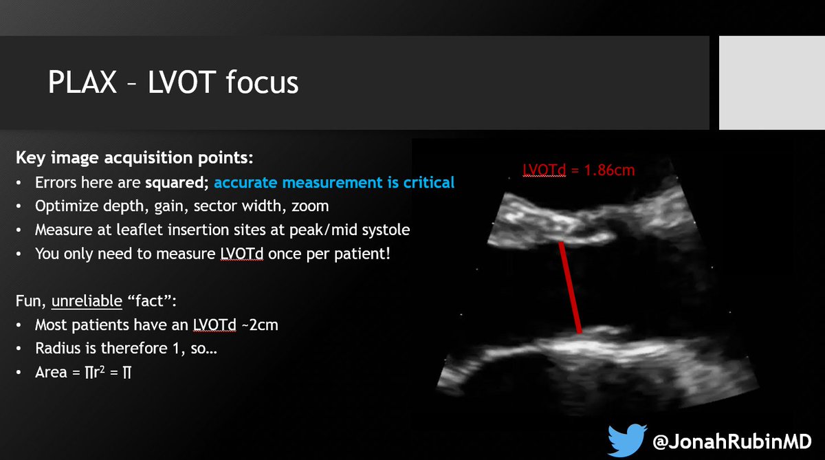
2⃣ Optimize the PLAX for the LVOTd. Sometimes I find that too much "optimization" actually makes it harder. Just know your options and do what works with your patient. ImageImage
3⃣ Understand Doppler physics. Specifically PWD for this one. Recognize how the angle of insonation AND the angle of flow relative to the probe, affects your measurements. ImageImage
Read 7 tweets
Sep 13
Greatly enjoyed launching our @MGH_PCCM @HarvardPulm didactic critical care echo curriculum with LV Assessment - Beyond the Eyeball!
Assessing LV function isn't as simple as it sounds.
Take home messages for POCUS learners below ⤵️:
#POCUS #Echofirst #Medtwitter #PCCMTwitter
👀The oft-invoked 'eyeball' method is a deceptive oversimplification. You can't suddenly accurately assess LV function just because you're told to!

You need to train your eye to look at other quantifiable measures of LF function - even if you won't actually measure them. 🧐
Sure - make your best guesstimate of EF. But also look at:
1⃣myocardial thickening
2⃣myocardial excursion
3⃣annular excursion/MAPSE
4⃣fractional shortening +/- fractional area change
5⃣EPSS
You don't have to actually measure these, but you can't 'see' them if you don't know them!
Read 7 tweets
Feb 7
🚨 A case of bactrim-associated ARDS!
BUT its backstory is one of sadness, selflessness, intrigue, sleuthing, clinical acumen, multidisciplinary collaboration, and most importantly, collaboration with patients and families themselves, led by @JennaMillerKC et al.
#Medtwitter
It's a tale and disease perhaps ultimately worthy of discussion in the medical historical annals of @AvrahamCooperMD @tony_breu @AdamRodmanMD.
But more importantly, worthy of widespread dissemination for broad awareness, rapid identification and management, and further research.
Our case, now published in @journal_CHEST, was cracked only based on the previous clinico-pathologic work of @JennaMillerKC, @JenniferGoldman, @MGHPathology Drs. Mari Mino-Kenudson, Angela Shih, & Martin Taylor, and @MGH_PCCM Drs. Taylor Thompson and @chivukula_raghu, and others.
Read 21 tweets
Feb 6
This harrowing, bone-chilling, terrifying, powerful account of a present day, ongoing, Holocaust while the United States participates in the Beijing Olympics eerily parallels its participation in Berlin's 1936. We must learn from our recent mistakes and act now.
This piece reads as play-by-play a reenactment of the stories I learned about my grandparents and friend's families in the Holocaust. Acceptance of a progressively more racist and genocidal state. "It's not that bad", "it can't get worse", "our whole life is here".
Conjured optimism about future improvement. Hushed tones about the present. Frequent disappearances. Sleeping in clothing anticipating a nighttime arrest, public humiliation, before being sent to a concentration camp.
Read 10 tweets
Feb 3
Thrilled to be at @accpchest Virtual Fellows Course! Will try to share bits and pearls as able on this thread (no guarantees!)

Thank you @meredithturetz @subanichandra @DanckersMD and all who created this!
Session 1: Systematic Approach to Interpreting CT Chest led by Dr. Suhail Raoof!
FOMO? See: Algorithmic Approach to the Interpretation of Diffuse Lung Disease on Chest CT Imaging in @journal_CHEST journal.chestnet.org/article/S0012-…
@journal_CHEST ⭐️ The secondary pulmonary lobule is your friend in teasing apart the lung parenchyma and defining where a disease process, anatomically, is taking place
⭐️ Hard to make them out? Start at the periphery
Read 26 tweets
Jan 6
The more you understand, the less you need to memorize. Take this 🫀 example:

Severe AR and MR are both defined by a regurgitant volume of 60mL. YET, another criterion, effective orifice area, must be 0.3cm^2 for AR but 0.4 for MR.

#Medtwitter #cardiotwitter #POCUS #Echofirst
I kept mixing up which is 0.3 and which is 0.4. We can keep trying to memorize this, or ask why? Why is the regurgitant volume the same, but the effective regurgitant orifice area (EROA) different?

Here's the pearl:
It's because AR occurs in diastole, MR occurs in systole, and diastole is longer than systole. Therefore, in diastole, a smaller EROA (0.3) can generally yield the same 60mL of regurgitation as a larger EROA (0.4) can during the shorter systole.
Read 5 tweets

Did Thread Reader help you today?

Support us! We are indie developers!


This site is made by just two indie developers on a laptop doing marketing, support and development! Read more about the story.

Become a Premium Member ($3/month or $30/year) and get exclusive features!

Become Premium

Don't want to be a Premium member but still want to support us?

Make a small donation by buying us coffee ($5) or help with server cost ($10)

Donate via Paypal

Or Donate anonymously using crypto!

Ethereum

0xfe58350B80634f60Fa6Dc149a72b4DFbc17D341E copy

Bitcoin

3ATGMxNzCUFzxpMCHL5sWSt4DVtS8UqXpi copy

Thank you for your support!

Follow Us on Twitter!

:(