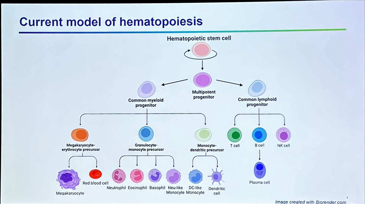
Best way to recognize pathological/neoplastic immunophenotypic changes is having a good grip on immunophynotypic variations in reactive/regenerative conditions… summarizing #FlowICCS22 plenary session 3 here👇🏻 🧵1/ #hemepath
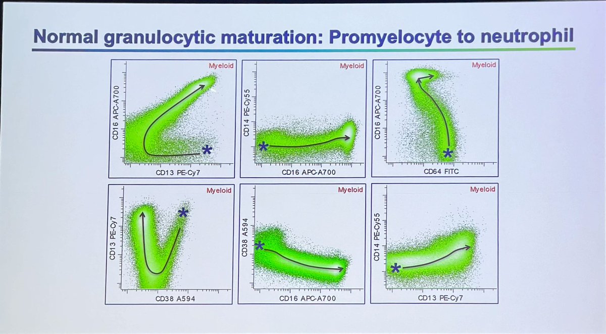
Normal granulocytic maturation patterns…. Plots follow maturation from promyelocyte (*) to neutrophils #flowiccs22#hemepath 2/
Here are phenotypic changes post GCSF therapy compared to Normal…. Don’t over-interpret this pattern as abnormal myeloid maturation #flowiccs22#hemepath 3/ promyelocyte (*) to neutrophil
Be aware of CD56 expression on normal myeloid progenitors, PDCs, and maturing myeloid and monocytic cells of individuals with #DownSyndrome don’t over-interpret as neoplastic changes #flowiccs22#hemepath 5/
• • •
Missing some Tweet in this thread? You can try to
force a refresh
Now that we know the answer is MDS-biTP53 let's see how we got to the answer...
I'll start from the beginning.. what exactly is this image that I showed you?
It's a whole genome view of using @bionano 's #OGM of a BM aspirate.. giving U a bird's-eye view of the chromosomes 1/
How do we interpret this? Easy!
normal cells have 2 copies of somatic chromosomes (1-22) one X & one Y if male, and 2 X if female.. (with some normal exceptions: i.e. men tend to lose their Y as they age... we think it's normal🤷♀️ or at least a very common somatic mosaicism) 2/
The chromosomes are on the X axis, their copy number is on Y axis! again, somatic chromosomes (1-22) should line up on 2 (red arrow) w/ roughly equal signal (above (blue) and below (red) the line).. here U can see parts of 4p (short arm), 4q(long arm) & 5q have 1 copy (loss) 3/
@AaronGoodman33 Typical cytomorphology is characterized by “blasts” with hand mirror (red arrow, cytoplasm to the side) or pearl necklace (green arrow, small cytoplasmic vacuoles around nucleus) appearance #bpdcn#hemepath#RareDisease#WHOHEME 1/
@AaronGoodman33 Skin and lymph node are most common extramedullary sites of involvement. Skin infiltrate is usually dermal with an appreciable “grenz zone” (uninvolved band b/w dermis and epidermis).. Lymph node typically has diffuse or paracortical involvement #hemepath#WHOHEME#BPDCN
One of the difficult aspects of #hemepath training is time management in a high-paced/demanding environment & the ability to multitask effectively while learning a difficult discipline. Every yr I offer our fellows helpful tips I’ve learned so far 🧵👇🏻More tips welcome,pls add 1/
Get rid of paper…
Don’t do anything twice
Take ownership of your cases… they are just as much yours as they are your faculty’s
Just another ordinary day at #hemepathMDA … 🤯
60-70 y/o woman
To all brave #hemepath aficionados how would you classify this case?
Poll and individual high quality images below. Pls comment if you’re feeling particularly brave today, let’s talk new #WHO 😎
The story of 1 day in the life of a #hematopathologist… I’m too exhausted to make a fancy educational thread but here are some amazing 🔬 pics for your viewing pleasure. I diagnosed all cases in one day 🤯 only at @MDAndersonNews Happy almost weekend people 🥂 #hemepath 🧵 1/n
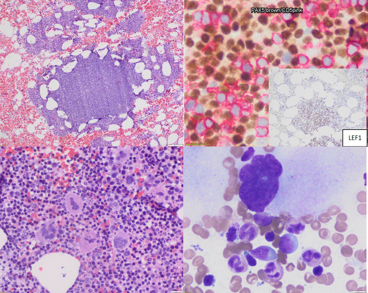
Myeloproliferative neoplasm w/ concurrent BCR::ABL1 and JAK2 V617F ..the megekaryocyte morphology is clue to something beyond CML #mpnsm#PathOutPic 2/
BPDCN with perfect so-called “hand mirror” (red) and “pearl necklace” (black) morphology #BPDCN#PathOutPic 3/
WT1 mutations are present in ~7% of de novo AML, are typically Lof mutation involving exons 7-9 of the gene. They frequently (~15%) co-occur w/ NPM1mut & have detrimental impact in this setting, shown by @AkEisfeld and colleagues in bit.ly/3eWQtXw@LeukemiaJnl
We studies a cohort of de novo NPM1-mut AML. 7% had concurrent WT1 mutations at baseline. 22% (15/67) relapsed; 4 (27%) with newly acquired Lof WT1-mut. Illustrated by @furudateken