The Neuroradiologist Profile picture
Nov 2, 2022 9 tweets 4 min read Read on X
Multiple Sclerosis - what to look for on MRI! A quick overview. First slide shows Jean-Martin Charcot, one of the fathers of Neurology (the painting is a bit outdated to say the least, but interesting from a historical point of view). #radres #neurorad #FOAMrad #FOAMed Image
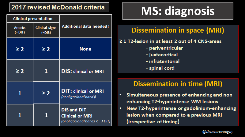
MS diagnosis is based on the McDonald criteria. To fulfill the diagnosis of MS, a patient must have evidence (clinical or radiological) of CNS-damage involving multiple CNS-regions (dissemination in space) and having occured at multiple moments in time (dissemination in time). Image
This 22-year old patient was referred for an MRI of the brain because of paresthesias in both lower legs. Lumbar MRI was normal. What do we make of those white matter lesions? Image
Let's localize those white matter lesions a bit better. The patient has periventricular, juxtacortical, infratentorial and spinal cord lesions. Image
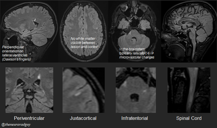
...and as such meets the MRI-requirements for dissemination in space. For dissemination in space on MRI we need at least 1 T2-hyperintense WM lesion in at least 2 out of 4 "MS-typical" regions. Image
On a first MRI in someone suspected of having MS, it's important to administer Gadolinium. Only so can we answer the quesion "dissemination in time". Simultaneous presence of enhancing and non-enhancing brain lesions = dissemiation in time. Image
Image
Alternatively, when previous MRI-exams are available, the presence of new T2-hyperintense white matter lesions (regardless of where) suffices to fulfill the criterium "dissemination in time". Image
And we finish with a nice summary of the McDonald criteria and the MRI-criteria for dissemation in space and time. Image

• • •

Missing some Tweet in this thread? You can try to force a refresh
 

Keep Current with The Neuroradiologist

The Neuroradiologist Profile picture

Stay in touch and get notified when new unrolls are available from this author!

Read all threads

This Thread may be Removed Anytime!

PDF

Twitter may remove this content at anytime! Save it as PDF for later use!

Try unrolling a thread yourself!

how to unroll video
  1. Follow @ThreadReaderApp to mention us!

  2. From a Twitter thread mention us with a keyword "unroll"
@threadreaderapp unroll

Practice here first or read more on our help page!

More from @theneuroradguy

Oct 31, 2022
Cystic supratentorial brain tumors in adults, the usual and the unusual. Non-exhaustive list of differential diagnostic considerations. The PA had a nice mural nodule, not shown on this figure. #radres #neurorad #FOAMrad #FOAMed #MedEd #RadEd
impossible to tell which is which on only T1-images with GD. Whenever I see an intra-axial tumor in an adult, I automatically consider GBM and metastases as they are the most frequent brain tumors in adults, so even when the tumor looks strange or atypical, they are a good bet.
Brain abcess and epidermoid cyst are pretty straightforward on diffusion-weighted images as their content is very diffusion restrictive. Brain abscess has a thick enhancing capsule, epidermoidcyst doesn't.
Read 8 tweets

Did Thread Reader help you today?

Support us! We are indie developers!


This site is made by just two indie developers on a laptop doing marketing, support and development! Read more about the story.

Become a Premium Member ($3/month or $30/year) and get exclusive features!

Become Premium

Don't want to be a Premium member but still want to support us?

Make a small donation by buying us coffee ($5) or help with server cost ($10)

Donate via Paypal

Or Donate anonymously using crypto!

Ethereum

0xfe58350B80634f60Fa6Dc149a72b4DFbc17D341E copy

Bitcoin

3ATGMxNzCUFzxpMCHL5sWSt4DVtS8UqXpi copy

Thank you for your support!

Follow Us!

:(