#POCUS quiz
You are performing physical examination in a patient with suspected fluid overload (plethoric IVC).
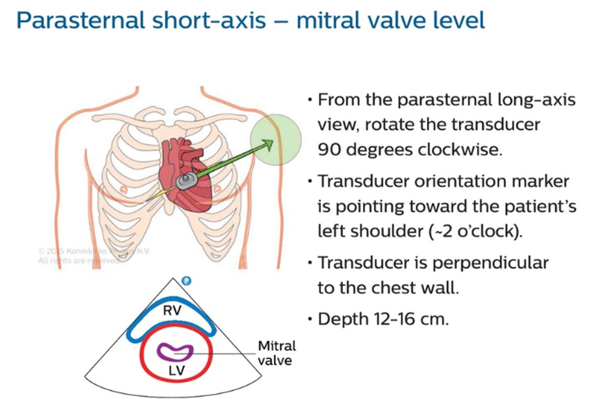
Parasternal short axis view demonstrates D-sign. But what's in the RV?
Apical view in thread.
#Nephrology #MedEd #FOAMcc
🔗 to source will be posted in a few hours.
You are performing physical examination in a patient with suspected fluid overload (plethoric IVC).
Parasternal short axis view demonstrates D-sign. But what's in the RV?
Apical view in thread.
#Nephrology #MedEd #FOAMcc
🔗 to source will be posted in a few hours.
Apical #POCUS
As our friends said, it's prominent moderator band in a patient with RV hypertrophy + prominent trabeculae.
Source article 🔗cvcasejournal.com/action/showPdf…
Source article 🔗cvcasejournal.com/action/showPdf…
• • •
Missing some Tweet in this thread? You can try to
force a refresh


