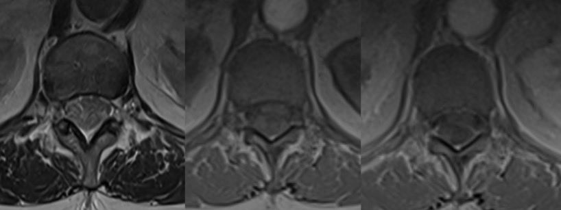
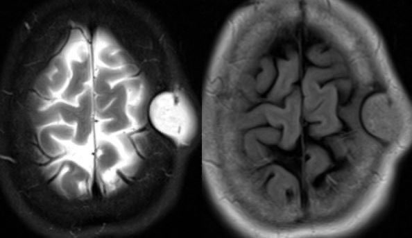
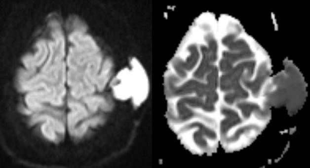
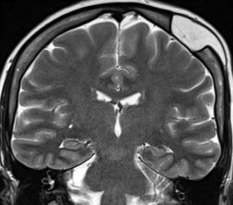
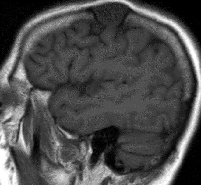
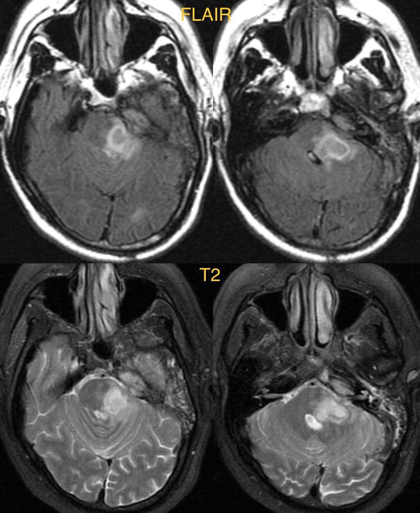
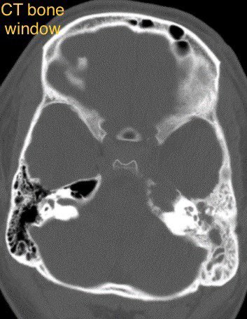
62 y/o M presents with signs of raised intracranial pressure. CT shows a hyper dense mass crossing the corpus callosum. On MR, the mass is hypointense on T2WI, restricting diffusion, and homogenously enhancing along the periventricular surface. #radtwitter#radres#neurotwitter
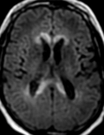
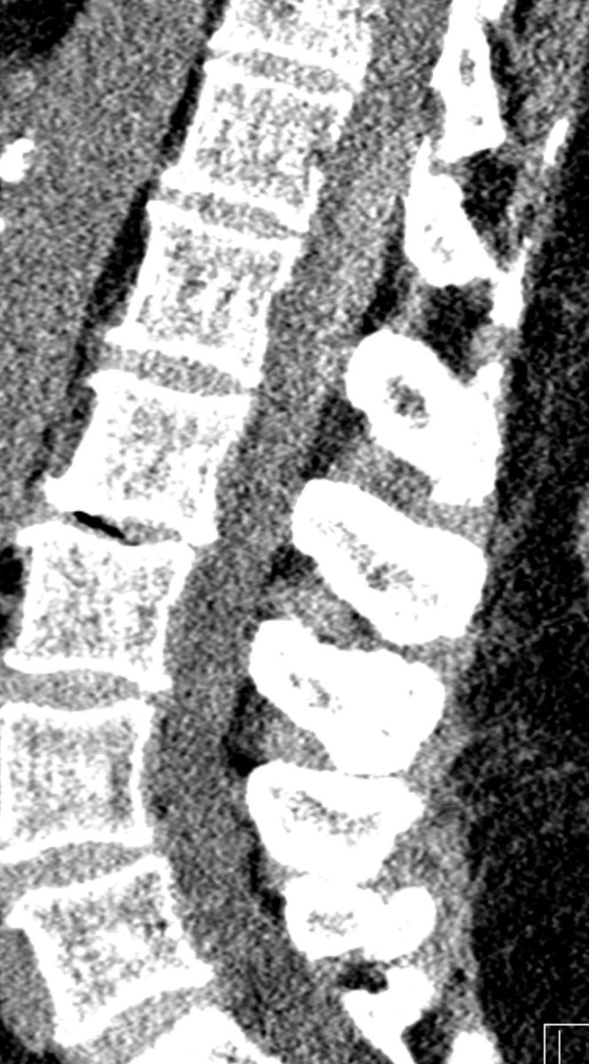
The corpus callosum is composed of very dense white matter tracks. Only aggressive tumors or lesions that effect the white matter cross the midline through the CC.
Diff Diagnosis for CC masses
High grade astrocytoma/Glioblastoma
Primary CNS lymphoma
Tumefactive Demyelination
Dx: Primary CNS lymphoma
PCNSL has a highly variable imaging appearance. Classically, it presents as a hyperdense mass with restricted diffusion and relatively hypointensity on T2WI due to hypercellularity. The mass enhances homogeneously and may cross the CC. #Neurosurgery
High grade astrocytoma and/or glioblastoma have more necrosis with heterogenous rather than homogenous enhancement.
Tumefactive demyelination has less mass effect relative to the size of the lesion, peripheral incomplete ring enhancement and marginal restricted diffusion. @RSNA
• • •
Missing some Tweet in this thread? You can try to
force a refresh
⭐️ Answer: Spontaneous spinal epidural hematoma (no clear risk factor in this case)
🔷CLINICAL: Abrupt onset of severe neck or back pain that can radiate into the extremities and commonly is followed by symptoms ranging from nerve root agitation to full neurologic impairment
🔷RISK FACTORS:
▶️Idiopathic (40-60%)
▶️Anticoagulant use
▶️AVM, AVF or other vascular malformation
▶️Underlying coagulopathy
▶️Tumor
▶️Pregnancy
❓ Possible explanation for cases without known risk factors: 🧠 🩸The internal epidural plexus that drains the abdomen and thorax is a low pressure, valveless system that may rupture when the pressure is increased from valsalva maneuvers ❓
🔷For glioblastoma we need to rely on many clinical and imaging features to distinguish (no one feature is specific enough to diagnose so we need to take the whole clinical and radiographic picture into account)
🔷Important features to consider:
▶️Recency of treatment
💡 Radiation necrosis is usually seen from 2 to 32 months after therapy, with 85% of cases occurring within 2 years. A new or worsening abnormality starting 3 years after completion of radiation therapy is very unlikely to be due to pure radiation necrosis. In the first 2–3 years, both tumor and radiation necrosis can occur
▶️Clinical symptoms (although both tumor progression and radiation necrosis can be symptomatic, we want to be careful calling tumor progression in a patient who is clinically doing well as this is devastating news for the patient and there are only limited options for therapy)
⭐️ Answer: petrous apicitis complicated by brainstem abscess
🔷Petrous apicitis can display the clinical triad of Gradenigo’s syndrome
1️⃣Otorrhea
2️⃣Cranial nerve 6 palsy
3️⃣Pain in distribution of trigeminal nerve
▶️Petrous apicitis usually occurs as a complication of otomastoiditis when the infection spreads to the skull base.
▶️The petrous apex is in close proximity to Dorello’s canal (where cranial nerve 6 runs through) and Meckel’s cave (where the trigeminal ganglion is located)
▶️CVT causes retrograde venous pressure leading to focal vasogenic edema
▶️Increased back pressure is characterized by dilated veins and petechial hemorrhage which can progress to large hematomas and ischemic neurological damage
🔷Two types of edema can develop:
1️⃣Vasogenic (from venous back pressure)
2️⃣Cytotoxic (ischemia)
🔷Risk factors:
💡 Up to 20% are idiopathic
1️⃣Trauma
2️⃣Tumor/malignancy (compression/invasion from meningioma)
3️⃣Infection
4️⃣Hormonal (pregnancy)
5️⃣Dehydration
🔷What is the most likely diagnosis in this 70 y/o F who lives with feral cats presenting w/ vomiting, diarrhea, leukocytosis, fever for 3 days and progressive decline in level of consciousness?
🔷CSF: initially normal, repeat a few days later ⬆️ WBC (lymphocyte predominant), ⬆️ Protein, normal glucose
🔷 South Central Asia and Southeast Asia appear to have the greatest number of cases
🔷Dogs are the most common reservoir 🐶 (bats in developed countries🦇)
🔷CLINICAL:
▶️Incubation period typically 3 weeks to 2 months (range 5 days to 6 months)
▶️Prodromal symptoms: fever, malaise, anxiety,
and itching at the inoculation site
💡Once in the body, the virus begins retrograde flow to extend to the dorsal root ganglion, which may correlate with neuropathic pain
▶️CNS manifestations: Mental status changes of excessive agitation and depression with hydrophobia and aerophobia