Wet mount and gram stain showing free hooklets of Echinococcus spp on cyst fluid from a 75 yr old F. Circled the hooklets in following pictures.

#Fortheloveofmicrobiology #clinicalmicrobiology #microrounds #IDpath #ASMClinMicro
#MicroTwitter #ClinMicro #microbiologypakistan
Echinococcosis or hydatid disease is caused by the larval stage of the dog tapeworm, Echinococcus granulosus. The definitive host for this disease is the dog or other canids and the intermediate hosts are cattle,sheep,pigs,goats or camels. Man is an accidental intermediate host
Hydatid disease in humans is potentially dangerous depending on the location of the cyst. Some cysts may remain undetected for many years until they become large enough to affect other organs. Symptoms are then of a space occupying lesion.
Lung cysts are usually asymptomatic until there is cough, shortness of breath or chest pain. Serious allergic sequelae, including anaphylactic shock, may occur if there is fluid leakage from the cyst in a patient previously sensitised by small fluid leaks into the circulation.
Imaging and serodiagnosis are the mainstay of diagnosis. Serological tests include Enzyme linked immunosorbent assay (ELISA), an indirect haemagglutination test and a complement fixation test. .
Microscopic examination of the cyst fluid to look for the characteristic protoscoleces which can be either invaginated or evaginated. The cyst fluid will also reveal free hooklets.

• • •

Missing some Tweet in this thread? You can try to force a refresh
 

Keep Current with fortheloveofmicrobiology

fortheloveofmicrobiology Profile picture

Stay in touch and get notified when new unrolls are available from this author!

Read all threads

This Thread may be Removed Anytime!

PDF

Twitter may remove this content at anytime! Save it as PDF for later use!

Try unrolling a thread yourself!

how to unroll video
  1. Follow @ThreadReaderApp to mention us!

  2. From a Twitter thread mention us with a keyword "unroll"
@threadreaderapp unroll

Practice here first or read more on our help page!

More from @fortheloveofmi2

Nov 20, 2022
India ink preparation of CSF from a patient with cryptococcal meningitis showing the budding yeast cells of C. neoformans surrounded by a characteristic wide gelatinous capsule. The yeasts also show narrow-base budding and characteristic variation in size.
The space occupied by the capsule shows as a clear space between the gray background of the ink particles and the refractile edge of the cell.
India ink/nigrosin stain is a negative, acid stain. This means that the dye easily gives up a hydrogen ion (proton) and the chromophore of the dye becomes negatively charged. Since the surface of most organims cells is negatively charged, the cell surface repels the dye.
Read 9 tweets
Nov 18, 2022
CSF received from a 12 year old child with Ewing sarcoma

India ink preparation showing rounded yeast cells surrounded by a halo, highly suggestive of Cryptococcus neoformans
#Fortheloveofmicrobiology #clinicalmicrobiology #mmidsp #microrounds #ASMClinMicro #microbiologypakistan Image
India ink stain, previously known as the Nigrosin stain, is a quick, low-resource method. It is widely used for the microscopic detection of cryptococci in CSF. Its a negative stain used to determine the organism’s cellular morphology.
In the India ink preparation, stain fills the background and the thick polysaccharide capsule does not take up the stain, resulting in an appearance of a refractile large white circle against a dark background, giving the appearance of a halo of light
Read 8 tweets
Feb 20, 2022
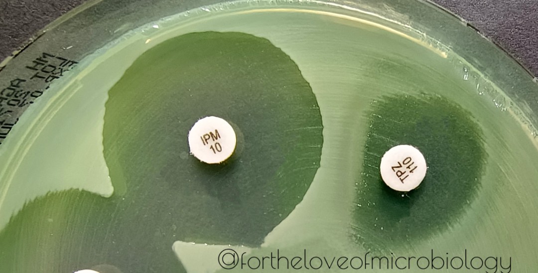
Many times when susceptibility testing is done for P. aeroginosa, a scenario similar to shown in the picture is encountered.
We see that TZP has produced a D shape on the IPM side. This might look very similar to the phenomenon seen in gram positive ICR strains. So what is it?
Published data says that this result is most likely due to
inducible expression of the P. aeruginosa AmpC beta-lactamase.
Certain enteric
(Serratia, Providencia, Citrobacter, Enterobacter, Morganella) and non-enteric organisms (P. aeruginosa, Aeromonas) can up-regulate expression of
their chromosomally-encoded ampC genes in response to sub-inhbitory concentrations of certain
b-lactam antibiotics
Read 10 tweets
Jul 8, 2021
E.coli, demonstrating the Eagle effect

The Eagle effect, Eagle phenomenon, or paradoxical zone phenomenon, named after Harry Eagle, originally referred to the paradoxically reduced antibacterial effect of penicillin at high doses.
Though recent usage generally refers to the relative lack of efficacy of beta lactam antibacterial drugs on infections having large numbers of bacteria. The former effect is paradoxical because the effectiveness of an antibiotic generally rises with increasing drug concentration.
In other words, the Eagle effect describes a phenomenon where decreased bactericidal activity of an organism is taking place despite of exposure to higher levels of an antibiotic.
Read 9 tweets

Did Thread Reader help you today?

Support us! We are indie developers!


This site is made by just two indie developers on a laptop doing marketing, support and development! Read more about the story.

Become a Premium Member ($3/month or $30/year) and get exclusive features!

Become Premium

Don't want to be a Premium member but still want to support us?

Make a small donation by buying us coffee ($5) or help with server cost ($10)

Donate via Paypal

Or Donate anonymously using crypto!

Ethereum

0xfe58350B80634f60Fa6Dc149a72b4DFbc17D341E copy

Bitcoin

3ATGMxNzCUFzxpMCHL5sWSt4DVtS8UqXpi copy

Thank you for your support!

Follow Us on Twitter!

:(