
Summiya Nizamuddin, Consultant Clinical Microbiologist, SKMCH&RC Pakistan 🔬🇵🇰
AKUH Alumnus, President MMIDSP


 Echinococcosis or hydatid disease is caused by the larval stage of the dog tapeworm, Echinococcus granulosus. The definitive host for this disease is the dog or other canids and the intermediate hosts are cattle,sheep,pigs,goats or camels. Man is an accidental intermediate host
 Yeast will grow on bacteriological media (sheep blood agar and chocolate agar).
 The space occupied by the capsule shows as a clear space between the gray background of the ink particles and the refractile edge of the cell.
 India ink stain, previously known as the Nigrosin stain, is a quick, low-resource method. It is widely used for the microscopic detection of cryptococci in CSF. Its a negative stain used to determine the organism’s cellular morphology.
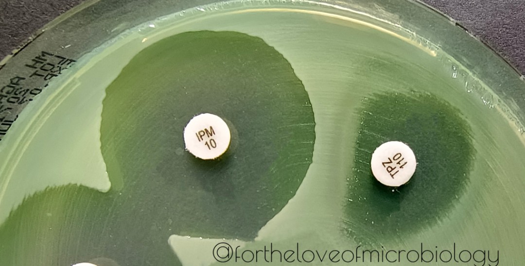 Published data says that this result is most likely due to
  Though recent usage generally refers to the relative lack of efficacy of beta lactam antibacterial drugs on infections having large numbers of bacteria. The former effect is paradoxical because the effectiveness of an antibiotic generally rises with increasing drug concentration.