NephroPOCUS Profile picture
Feb 1 4 tweets 3 min read
A 58-year-old woman with no known comorbidities presents with progressive fatigue and shortness of breath x several months. Noted to have bilateral pedal edema; BNP 2,473 pg/mL.
#echofirst 👇❓
Answer and 🔗 to source in thread.
#POCUS #MedEd #FOAMcc
Left atrial myxoma -> pulmonary hypertension (RVSP 93 mmHg) -> RV dysfunction (Note obvious RV enlargement ☝️
cvcasejournal.com/article/S2468-…
PLAX (same case)
Typical locations of various cardiac tumors and masses.
May help formulating differential diagnosis during #POCUS #echofirst
Image from Tyebally, et al. JACC 2020.

• • •

Missing some Tweet in this thread? You can try to force a refresh
 

Keep Current with NephroPOCUS

NephroPOCUS Profile picture

Stay in touch and get notified when new unrolls are available from this author!

Read all threads

This Thread may be Removed Anytime!

PDF

Twitter may remove this content at anytime! Save it as PDF for later use!

Try unrolling a thread yourself!

how to unroll video
  1. Follow @ThreadReaderApp to mention us!

  2. From a Twitter thread mention us with a keyword "unroll"
@threadreaderapp unroll

Practice here first or read more on our help page!

More from @NephroP

Feb 1
After yesterday's #POCUS quiz, it's time to reshare these cardiac tamponade infographics.
Courtesy of @ACEP_EUS
🔗acep.org/emultrasound/s…
Set of 3
See 🧵for the rest
#Nephpearls #MedEd #FOAMcc Image
Pulsus paradoxus #echofirst Image
Hepatic vein waveform changes
#POCUS #VExUS Image
Read 5 tweets
Jan 30
Another set of cardiac #POCUS #anatomy illustrations. 🧵
#Nephpearls #FOAMed
Source: sciencedirect.com/science/articl…
1⃣ Parasternal long axis
2⃣ Parasternal short axis aortic valve level
#POCUS
3⃣ Apical 4-chamber view #POCUS
Read 7 tweets
Jan 27
#MedEd image of the day. What's wrong with the 🫀?
Clinical: A woman in 40s with h/o severe mitral stenosis secondary to rheumatic heart disease & Afib presented with worsening dyspnea, orthopnea, & chest pain x 7 months
See thread for #echofirst images and source/answer.
#POCUS Image
Apical 4-chamber view showing severe right atrial enlargement and annular dilatation of the tricuspid valve during systole.
Severe TR as expected.
Read 5 tweets
Dec 28, 2022
#POCUS puzzle 🧩of the day. Can you put together the pieces and arrive at an unifying diagnosis? Go through the thread 🧵below.
Context: #dialysis patient, physical examination #FOAMed #Nephrology #MedEd

Image No. 1 👇
Color #POCUS
Transverse #POCUS , IVC trace down
Read 8 tweets
Dec 6, 2022
#POCUS quiz
You are performing physical examination in a patient with suspected fluid overload (plethoric IVC).
Parasternal short axis view demonstrates D-sign. But what's in the RV?
Apical view in thread.
#Nephrology #MedEd #FOAMcc
🔗 to source will be posted in a few hours.
Apical #POCUS
As our friends said, it's prominent moderator band in a patient with RV hypertrophy + prominent trabeculae.
Source article 🔗cvcasejournal.com/action/showPdf…
Read 4 tweets
Nov 29, 2022
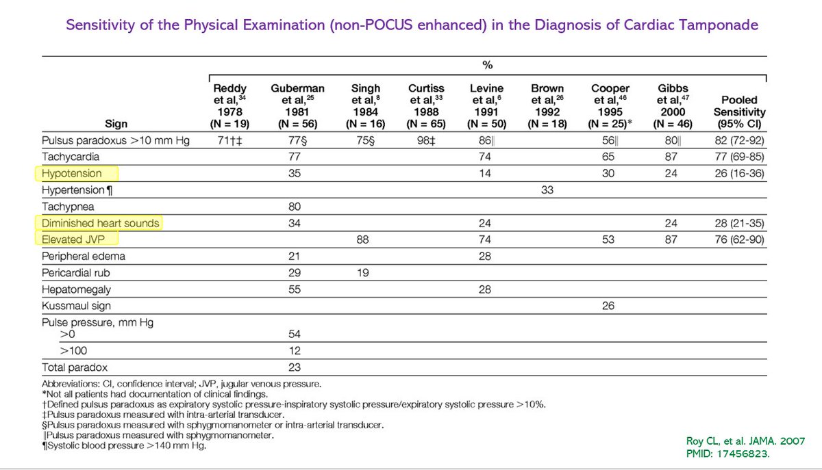
#POCUS #tamponade #MedEd
Short axis view and 🔗to source in thread.
👆image shows parasternal long axis view with diastolic collapse of the right ventricle.
👇short axis view at the aortic valve level showing right atrial systolic collapse (arrowhead) and RV diastolic collapse (arrow).
Source: cvcasejournal.com/article/S2468-…
#POCUS #echofirst #MedEd
Conventional physical exam!
Read 5 tweets

Did Thread Reader help you today?

Support us! We are indie developers!


This site is made by just two indie developers on a laptop doing marketing, support and development! Read more about the story.

Become a Premium Member ($3/month or $30/year) and get exclusive features!

Become Premium

Don't want to be a Premium member but still want to support us?

Make a small donation by buying us coffee ($5) or help with server cost ($10)

Donate via Paypal

Or Donate anonymously using crypto!

Ethereum

0xfe58350B80634f60Fa6Dc149a72b4DFbc17D341E copy

Bitcoin

3ATGMxNzCUFzxpMCHL5sWSt4DVtS8UqXpi copy

Thank you for your support!

Follow Us on Twitter!

:(