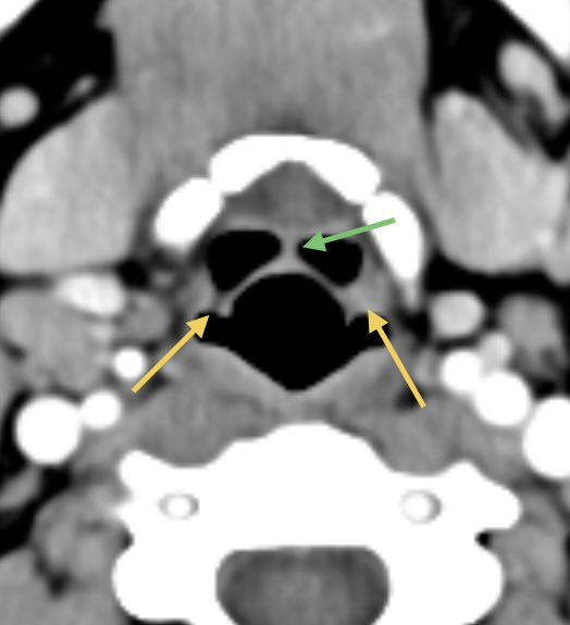
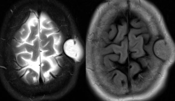
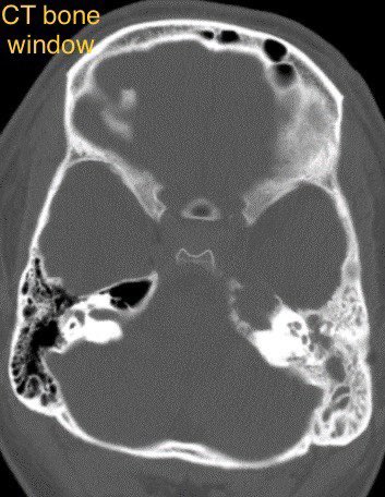
Congenital: imperforate membrane, incomplete canalization, or atresia leads to tears and mucus accumulation causing a dacryocystocele, if infected/inflamed, use the term dacryocystitis
Adults: acquired from any cause of obstruction
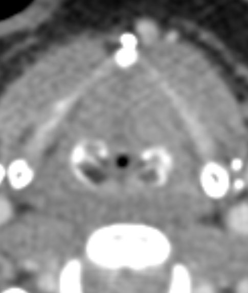
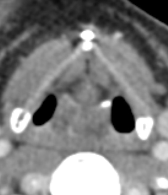
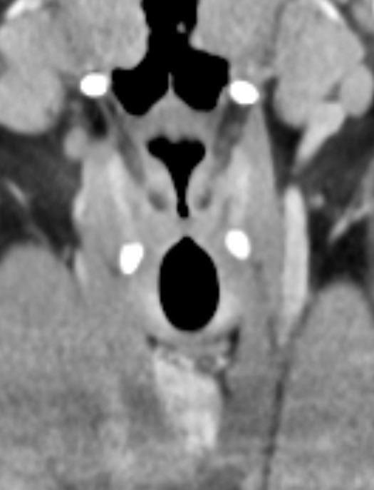
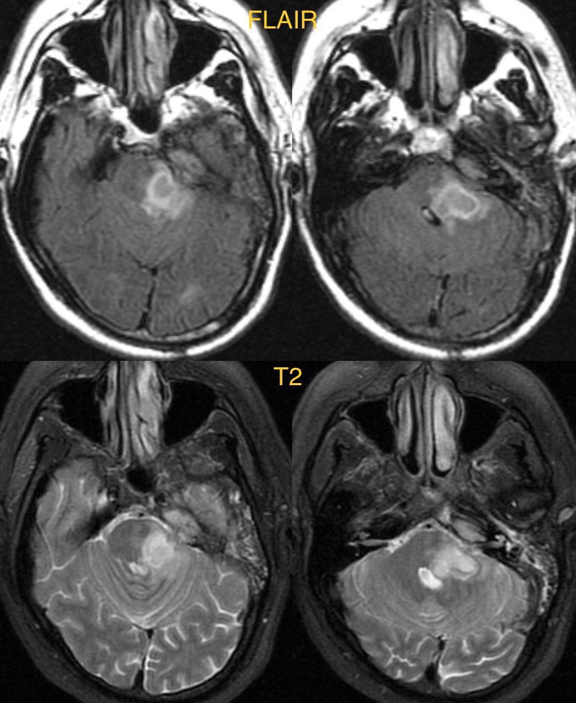
Imaging: often obtained to evaluate for orbital cellulitis
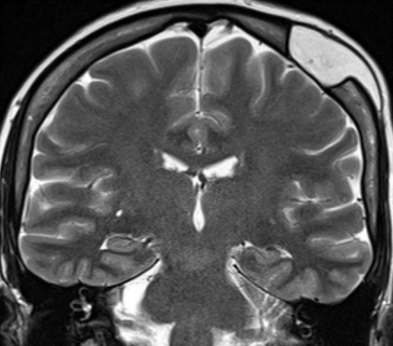
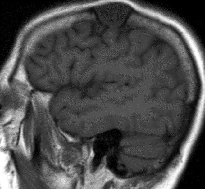
Look for a well defined cystic lesion at the medial canthus with continuity with the nasolacrimal duct
When reporting, discuss the extent of the lesion, laterality, and if there are signs of infection 🧠
• • •
Missing some Tweet in this thread? You can try to
force a refresh
🔷For glioblastoma we need to rely on many clinical and imaging features to distinguish (no one feature is specific enough to diagnose so we need to take the whole clinical and radiographic picture into account)
🔷Important features to consider:
▶️Recency of treatment
💡 Radiation necrosis is usually seen from 2 to 32 months after therapy, with 85% of cases occurring within 2 years. A new or worsening abnormality starting 3 years after completion of radiation therapy is very unlikely to be due to pure radiation necrosis. In the first 2–3 years, both tumor and radiation necrosis can occur
▶️Clinical symptoms (although both tumor progression and radiation necrosis can be symptomatic, we want to be careful calling tumor progression in a patient who is clinically doing well as this is devastating news for the patient and there are only limited options for therapy)
⭐️ Answer: petrous apicitis complicated by brainstem abscess
🔷Petrous apicitis can display the clinical triad of Gradenigo’s syndrome
1️⃣Otorrhea
2️⃣Cranial nerve 6 palsy
3️⃣Pain in distribution of trigeminal nerve
▶️Petrous apicitis usually occurs as a complication of otomastoiditis when the infection spreads to the skull base.
▶️The petrous apex is in close proximity to Dorello’s canal (where cranial nerve 6 runs through) and Meckel’s cave (where the trigeminal ganglion is located)
▶️CVT causes retrograde venous pressure leading to focal vasogenic edema
▶️Increased back pressure is characterized by dilated veins and petechial hemorrhage which can progress to large hematomas and ischemic neurological damage
🔷Two types of edema can develop:
1️⃣Vasogenic (from venous back pressure)
2️⃣Cytotoxic (ischemia)
🔷Risk factors:
💡 Up to 20% are idiopathic
1️⃣Trauma
2️⃣Tumor/malignancy (compression/invasion from meningioma)
3️⃣Infection
4️⃣Hormonal (pregnancy)
5️⃣Dehydration
🔷What is the most likely diagnosis in this 70 y/o F who lives with feral cats presenting w/ vomiting, diarrhea, leukocytosis, fever for 3 days and progressive decline in level of consciousness?
🔷CSF: initially normal, repeat a few days later ⬆️ WBC (lymphocyte predominant), ⬆️ Protein, normal glucose
🔷 South Central Asia and Southeast Asia appear to have the greatest number of cases
🔷Dogs are the most common reservoir 🐶 (bats in developed countries🦇)
🔷CLINICAL:
▶️Incubation period typically 3 weeks to 2 months (range 5 days to 6 months)
▶️Prodromal symptoms: fever, malaise, anxiety,
and itching at the inoculation site
💡Once in the body, the virus begins retrograde flow to extend to the dorsal root ganglion, which may correlate with neuropathic pain
▶️CNS manifestations: Mental status changes of excessive agitation and depression with hydrophobia and aerophobia
🔷35 y/o F w/ history of Li Fraumeni syndrome presents w/ intermittent left sided weakness and pain. The feeling is of heaviness, difficulty w/ grip, dropping objects and frequent falls. Episodes last for weeks to months. What is your diagnosis? 🧠
🎉Congrats to all the rad fellow matches today!!! 🍾
⭐️ Answer: Tumefactive demyelination (MS in this case)
🔷Imaging in this case is specific enough to diagnosis with confidence but we need extra caution in patients with genetic predisposition to malignancy
💡 In cases where imaging or clinical picture are atypical or discordant for demyelination (especially if there is a genetic disorder), it is best to refer to neurology for proper work up, possible trial of steroids and short imaging follow up
🚩 Be suspicious against MS if your patient has:
1️⃣Systemic symptoms (fever, weight loss, joint or skin symptoms, etc.)
2️⃣Seizures, hearing loss, meningitis signs, movement disorder, aphasia
3️⃣”Family history”
4️⃣Age (<20 or >50)
5️⃣Lesions are symmetric
6️⃣Hemorrhage or dense on CT
7️⃣Diffusion restriction other than leading edge
8️⃣Strokes
9️⃣Cysts
🔟Cortical infiltration