#ContinuumCase, plot twist edition:
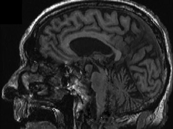
A 28 year old PGY2 neurology resident is asked by her attending to review 2 scans and determine the diagnosis: Alzheimer disease (AD) or dementia with Lewy bodies (DLB).
What should she say, #neurotwitter?🧵
A 28 year old PGY2 neurology resident is asked by her attending to review 2 scans and determine the diagnosis: Alzheimer disease (AD) or dementia with Lewy bodies (DLB).
What should she say, #neurotwitter?🧵

How would you respond?
Her response:
“Dementia is a clinical diagnosis supported by imaging, CSF, and neuropathological biomarkers. But, the disproportionate hippocampal atrophy in Patient B supports a diagnosis of AD.”
“Dementia is a clinical diagnosis supported by imaging, CSF, and neuropathological biomarkers. But, the disproportionate hippocampal atrophy in Patient B supports a diagnosis of AD.”

Undeterred, the attending asks her to review the FDG PET images as well.
(little does he know, she subscribes to Continuum)
(little does he know, she subscribes to Continuum)

Her response:
“Well, the frontal and lateral parietal hypometabolism in Patient B’s study again favors AD. Furthermore, the occipitoparietal hypometabolism in Patient A’s scan is suggestive of DLB.”
The attending starts to get nervous and break out in a sweat.
“Well, the frontal and lateral parietal hypometabolism in Patient B’s study again favors AD. Furthermore, the occipitoparietal hypometabolism in Patient A’s scan is suggestive of DLB.”
The attending starts to get nervous and break out in a sweat.
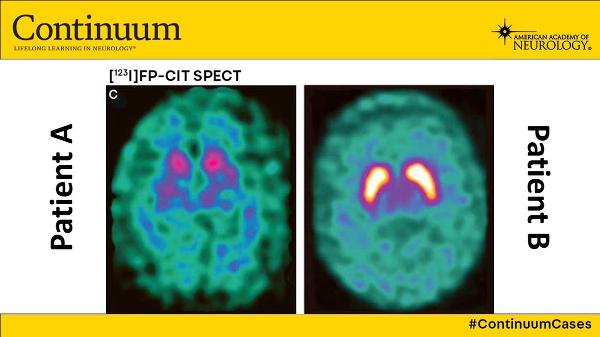
Attending: “Well how about THESE images?”
Resident: “This is clearly a dopamine transporter single photon emission computed tomogram (DAT SPECT) indicating symmetrically reduced putaminal uptake. Again, favors DLB in Patient A.”
✋
🎤
Resident: “This is clearly a dopamine transporter single photon emission computed tomogram (DAT SPECT) indicating symmetrically reduced putaminal uptake. Again, favors DLB in Patient A.”
✋
🎤

For a complete review of Neuroimaging in Dementia, check out this excellent article from Drs. Shannon Risacher (@RisacherShannon) and Liana Apostolova (@ApostolovaLiana)! @IU_Health
journals.lww.com/continuum/Full…
journals.lww.com/continuum/Full…
• • •
Missing some Tweet in this thread? You can try to
force a refresh


