One of the most important diagnostic tests in Cardiology to interpret is the EKG.

Here are my thoughts and notes. Will continue to this thread. Let me know what you think!

Thread #8: Right Bundle Branch Block (RBBB)

#arjuncardiology #medtwitter #CardioTwitter #MedEd #IMG
Ventricular Conduction:
- Normal electrical stimulus reaches ventricles from the atria through the AV node & His-Purkinje systems
- First part of heart to be depolarized is the left-side of the septum; then spreads to RV and LV by right & left bundles
- Normal QRS < 0.10 sec
RBBB:
- 1st phase of depolarization: Left side of septum is stimulated first (branch of left bundle); on a normal ECG produces a septal r-wave in V1 and small septal q-wave in V6. No impact with RBBB.

- 2nd phase: Simultaneous depolarization of LV and RV. No impact with RBBB.
RBBB:
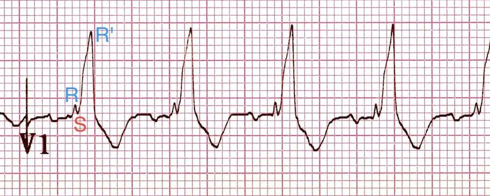
- 3rd phase: Delayed RV depolarization produces a third phase of ventricular stimulation. V1 (right-sided chest leads) records this phase of ventricular stimulation as a (+) wide deflection (R' wave).
- Will see a wide negative S-wave deflection in left-sided chest leads
Complete and Incomplete RBBB:
- Complete: QRS > 0.12 seconds in duration w/ rSR' in lead V1 and qRS in V6

- Incomplete: Same QRS patterns, but duration is between 0.10 - 0.12 seconds
RBBB:
- May not have underlying heart disorder
- Can be seen with ASD, COPD with pulmonary hypertension, pulmonary stenosis, cardiomyopathy, and CAD
- No specific treatment, can be permanent or transient
Thank you to this amazing website for the graphics: litfl.com/right-bundle-b…

Also this is another great video:

Let me know what you think and stay tuned for left bundle branch block next!

• • •

Missing some Tweet in this thread? You can try to force a refresh
 

Keep Current with Arjun Khadilkar, MD

Arjun Khadilkar, MD Profile picture

Stay in touch and get notified when new unrolls are available from this author!

Read all threads

This Thread may be Removed Anytime!

PDF

Twitter may remove this content at anytime! Save it as PDF for later use!

Try unrolling a thread yourself!

how to unroll video
  1. Follow @ThreadReaderApp to mention us!

  2. From a Twitter thread mention us with a keyword "unroll"
@threadreaderapp unroll

Practice here first or read more on our help page!

More from @akhadilkarMD

Mar 16
One of the most important diagnostic tests in Cardiology to interpret is the EKG.

Here are my thoughts and notes. Will continue to this thread. Let me know what you think!

Thread #9: Left Bundle Branch Block (LBBB)

#arjuncardiology #medtwitter #CardioTwitter #MedEd #IMG
LBBB:
- Similar to a RBBB, produces a wide QRS and affects the early phase of depolarization
- Septum will depolarize from (right to left; instead of normal left to right).
- Will see the loss of septal r-wave in V1 and septal q-wave in V6 ImageImage
LBBB:
- V1: Negative QRS complex b/c the LV is still electrically predominant (initial depolarization is negative and remains negative in the right-sided chest lead) (W-shape)
- V6: Entirely positive R-wave ('M'- Pattern) Image
Read 6 tweets
Mar 3
One of the most important diagnostic tests in Cardiology to interpret is the EKG.

Here are my thoughts and notes. Will continue to this thread. Let me know what you think!

Thread #7: Ventricular Hypertrophy

#arjuncardiology #medtwitter #CardioTwitter #MedEd #IMG
Atrial and Ventricular Enlargement:
- Both dilation & hypertrophy usually result in chronic pressure and volume overload on the heart muscle

- Pathological hypertrophy & dilation are often accompanied by fibrosis (scarring); can lead to arrhythmias and heart failure.
Right Ventricular Hypertrophy:
- Right chest leads show tall R-waves
- R-wave > S-wave in V1 is suggestive; not diagnostic of RVH
- Can see right-axis deviation and T-wave inversions in the right & mid-precordial leads
- RV hypertrophy can lead to variations in repolarization
Read 9 tweets
Mar 2
One of the most important diagnostic tests in Cardiology to interpret is the EKG.

Here are my thoughts and notes. Will continue to this thread. Let me know what you think!

Thread #6: Atrial Abnormality

#arjuncardiology #medtwitter #CardioTwitter #MedEd #IMG
Atrial and Ventricular Enlargement:
- Both dilation & hypertrophy usually result in chronic pressure and volume overload on the heart muscle

- Pathological hypertrophy & dilation are often accompanied by fibrosis (scarring); can lead to arrhythmias and heart failure.
Right Atrial Abnormality (RAA):
- Overload of RA (dilation/ hypertrophy) may increase P-wave voltage

- Normal P-wave < 2.5 mm amplitude and < 0.12 seconds in width

- Tall, narrow P-waves: characteristic of RAA and can be best seen in II, III, aVF
Read 7 tweets
Feb 6
One of the most important diagnostic tests in Cardiology to interpret is the EKG.

Here are my thoughts and notes. Will continue to this thread. Let me know what you think!

Thread #5: Axis Deviation

#arjuncardiology #medtwitter #CardioTwitter #MedEd #IMG
QRS Axis:
- General direction in the frontal plane towards which the QRS complex is predominantly pointed

- General rule: Mean QRS points mid-way b/w any 2 leads that show tall R-wave of equal height

- If depolarization is perpendicular to any lead, will see biphasic complex
Axis Deviation:
- For most people, the axis lies between -30 and + 100 degrees

- Left axis: < -30 degrees, lead II rS pattern (S-wave deeper than R-wave is tall)

- Right axis: > +100 degrees (R-wave in III > II)
Read 6 tweets
Feb 6
One of the most important diagnostic tests in Cardiology to interpret is the EKG.

Here are my thoughts and notes. Will continue to this thread. Let me know what you think!

Thread #4: ECG Segments

#arjuncardiology #medtwitter #CardioTwitter #MedEd #IMG
General Principles:
- Positive deflection: wave of depolarization towards positive pole of that lead
- Negative deflection: wave of depolarization towards negative pole of that lead
- Biphasic deflection: wave of depolarization is perpendicular to a lead
Normal Sinus P-wave:
- Atrial depolarization that marks spontaneous depolarization of pacemakers cells in the right atrium
- Should be negative P-wave in aVR and upright in lead II
- Can communicate 'sinus rhythm with 1:1 AV conduction'
Read 7 tweets
Feb 5
One of the most important diagnostic tests in Cardiology to interpret is the EKG.

Here are my thoughts and notes. Will continue to this thread. Let me know what you think!

Thread #3: ECG Leads

#arjuncardiology #medtwitter #CardioTwitter #MedEd #IMG
ECG Leads:
- Body act as a conductor of electricity; the recording electrodes in the arms, legs, and chest wall show the differences in voltage (potential) among electrodes
- Different views of the same event leads to different ECG patterns
ECG Limb Leads:
- 6 Limb leads (extremity leads) and 6 chest (precordial)
- 3 bipolar limb leads: I, II, III
- 3 augmented unipolar: aVR, aVL, and aVF
- 6 precordial leads: V1-V6
Read 7 tweets

Did Thread Reader help you today?

Support us! We are indie developers!


This site is made by just two indie developers on a laptop doing marketing, support and development! Read more about the story.

Become a Premium Member ($3/month or $30/year) and get exclusive features!

Become Premium

Don't want to be a Premium member but still want to support us?

Make a small donation by buying us coffee ($5) or help with server cost ($10)

Donate via Paypal

Or Donate anonymously using crypto!

Ethereum

0xfe58350B80634f60Fa6Dc149a72b4DFbc17D341E copy

Bitcoin

3ATGMxNzCUFzxpMCHL5sWSt4DVtS8UqXpi copy

Thank you for your support!

Follow Us on Twitter!

:(