One of the most important diagnostic tests in Cardiology to interpret is the EKG.

Here are my thoughts and notes. Will continue to this thread. Let me know what you think!

Thread #11: Myocardial Ischemia

#arjuncardiology #medtwitter #CardioTwitter #MedEd #IMG
Myocardial Ischemia:
- One of the most important things to evaluate on EKG
- If severe narrowing/complete blockage of a coronary artery causes blood flow to become adequate, ischemia of the heart muscle develops
- Can be transient (angina pectoris) or more severe (necrosis & MI)
Myocardial Ischemia
- LV consists of an outer layer (epicardium/sub-epicardium) and inner layer (sub-endocardium)
- Can have limit of ischemia to the inner layer or can affect the entire thickness of the ventricular wall (transmural ischemia)
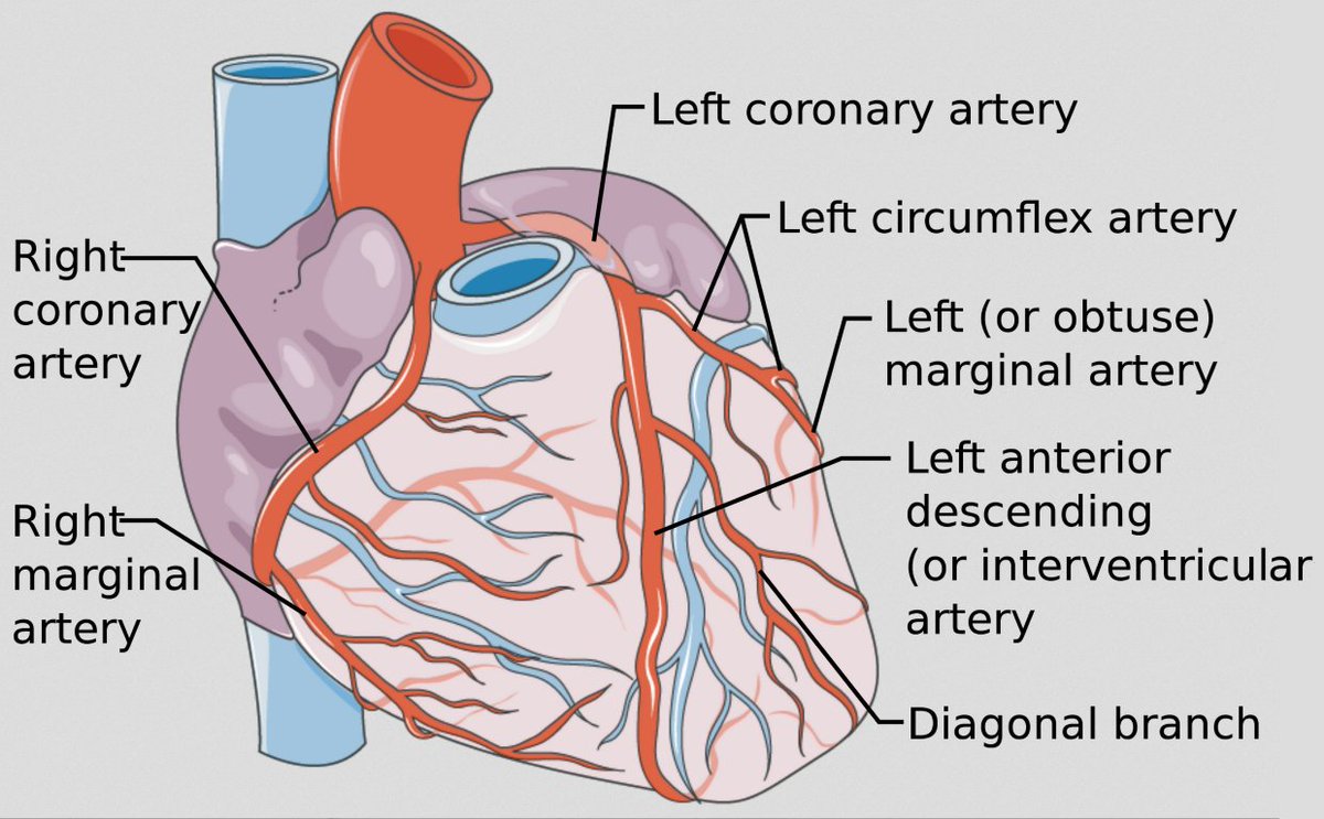
Myocardial Blood Supply
- RCA supplies both inferior (diaphragmatic) portion of the heart and RV
- Left main divides into the left anterior descending artery (LAD), which supplies the ventricular septum and large part of free LV wall, and LCx (lateral wall of the LV)
ST Elevation/Transmural Ischemia
- Ischemia & ultimately necrosis of a portion of the LV free wall
- Most patients w/ acute MI have underlying atherosclerotic disease
- Occlusion of coronary artery with a ruptured plaque leads to coagulation cascade (fibrin & platelets)
ST Elevation/Transmural Ischemia
- Other factors for acute STEMI: cocaine, coronary artery dissection, coronary artery emboli
- Acute phase: ST elevations, hyper-acute T-waves in multiple leads
- Evolving phase: Hours/days later with deep T-wave inversions replacing ST elevation
ST Elevation/Transmural Ischemia
- Anterior MI: infarct of anterior/lateral wall of LV
- Inferior MI: infarct of inferior wall of LV
- ST segment (& reciprocal) changes generally seen within minutes of blood flow occlusion
- Reperfusion therapy can decrease ST elevations
Thanks to these amazing website for the wonderful graphics:

litfl.com/myocardial-isc…
grepmed.com/images/3929/ek…
ccasociety.org/education/echo…

Stay tuned for the next thread on Q-waves!

• • •

Missing some Tweet in this thread? You can try to force a refresh
 

Keep Current with Arjun Khadilkar, MD

Arjun Khadilkar, MD Profile picture

Stay in touch and get notified when new unrolls are available from this author!

Read all threads

This Thread may be Removed Anytime!

PDF

Twitter may remove this content at anytime! Save it as PDF for later use!

Try unrolling a thread yourself!

how to unroll video
  1. Follow @ThreadReaderApp to mention us!

  2. From a Twitter thread mention us with a keyword "unroll"
@threadreaderapp unroll

Practice here first or read more on our help page!

More from @akhadilkarMD

Mar 17
One of the most important diagnostic tests in Cardiology to interpret is the EKG.

Here are my thoughts and notes. Will continue to this thread. Let me know what you think!

Thread #10: Fascicular Blocks

#arjuncardiology #medtwitter #CardioTwitter #MedEd #IMG
Fascicular Blocks:
- Left bundle branch system: sub-divided into an anterior & posterior fascicle.

- Hemi-block does not widen the QRS complex markedly (compared to a RBBB or LBBB)
Left Anterior Fascicular Block (LAFB):
- Diagnosed by finding of a left axis deviation (-45 degrees or more negative)
- Delayed activation of more superior & leftward position of the LV
- Isolated finding is non-specific; can be seen w/ HTN, AV disease, CAD, and aging
Read 9 tweets
Mar 16
One of the most important diagnostic tests in Cardiology to interpret is the EKG.

Here are my thoughts and notes. Will continue to this thread. Let me know what you think!

Thread #9: Left Bundle Branch Block (LBBB)

#arjuncardiology #medtwitter #CardioTwitter #MedEd #IMG
LBBB:
- Similar to a RBBB, produces a wide QRS and affects the early phase of depolarization
- Septum will depolarize from (right to left; instead of normal left to right).
- Will see the loss of septal r-wave in V1 and septal q-wave in V6
LBBB:
- V1: Negative QRS complex b/c the LV is still electrically predominant (initial depolarization is negative and remains negative in the right-sided chest lead) (W-shape)
- V6: Entirely positive R-wave ('M'- Pattern)
Read 6 tweets
Mar 15
One of the most important diagnostic tests in Cardiology to interpret is the EKG.

Here are my thoughts and notes. Will continue to this thread. Let me know what you think!

Thread #8: Right Bundle Branch Block (RBBB)

#arjuncardiology #medtwitter #CardioTwitter #MedEd #IMG
Ventricular Conduction:
- Normal electrical stimulus reaches ventricles from the atria through the AV node & His-Purkinje systems
- First part of heart to be depolarized is the left-side of the septum; then spreads to RV and LV by right & left bundles
- Normal QRS < 0.10 sec
RBBB:
- 1st phase of depolarization: Left side of septum is stimulated first (branch of left bundle); on a normal ECG produces a septal r-wave in V1 and small septal q-wave in V6. No impact with RBBB.

- 2nd phase: Simultaneous depolarization of LV and RV. No impact with RBBB.
Read 7 tweets
Mar 3
One of the most important diagnostic tests in Cardiology to interpret is the EKG.

Here are my thoughts and notes. Will continue to this thread. Let me know what you think!

Thread #7: Ventricular Hypertrophy

#arjuncardiology #medtwitter #CardioTwitter #MedEd #IMG
Atrial and Ventricular Enlargement:
- Both dilation & hypertrophy usually result in chronic pressure and volume overload on the heart muscle

- Pathological hypertrophy & dilation are often accompanied by fibrosis (scarring); can lead to arrhythmias and heart failure.
Right Ventricular Hypertrophy:
- Right chest leads show tall R-waves
- R-wave > S-wave in V1 is suggestive; not diagnostic of RVH
- Can see right-axis deviation and T-wave inversions in the right & mid-precordial leads
- RV hypertrophy can lead to variations in repolarization
Read 9 tweets
Mar 2
One of the most important diagnostic tests in Cardiology to interpret is the EKG.

Here are my thoughts and notes. Will continue to this thread. Let me know what you think!

Thread #6: Atrial Abnormality

#arjuncardiology #medtwitter #CardioTwitter #MedEd #IMG
Atrial and Ventricular Enlargement:
- Both dilation & hypertrophy usually result in chronic pressure and volume overload on the heart muscle

- Pathological hypertrophy & dilation are often accompanied by fibrosis (scarring); can lead to arrhythmias and heart failure.
Right Atrial Abnormality (RAA):
- Overload of RA (dilation/ hypertrophy) may increase P-wave voltage

- Normal P-wave < 2.5 mm amplitude and < 0.12 seconds in width

- Tall, narrow P-waves: characteristic of RAA and can be best seen in II, III, aVF
Read 7 tweets
Feb 6
One of the most important diagnostic tests in Cardiology to interpret is the EKG.

Here are my thoughts and notes. Will continue to this thread. Let me know what you think!

Thread #5: Axis Deviation

#arjuncardiology #medtwitter #CardioTwitter #MedEd #IMG
QRS Axis:
- General direction in the frontal plane towards which the QRS complex is predominantly pointed

- General rule: Mean QRS points mid-way b/w any 2 leads that show tall R-wave of equal height

- If depolarization is perpendicular to any lead, will see biphasic complex
Axis Deviation:
- For most people, the axis lies between -30 and + 100 degrees

- Left axis: < -30 degrees, lead II rS pattern (S-wave deeper than R-wave is tall)

- Right axis: > +100 degrees (R-wave in III > II)
Read 6 tweets

Did Thread Reader help you today?

Support us! We are indie developers!


This site is made by just two indie developers on a laptop doing marketing, support and development! Read more about the story.

Become a Premium Member ($3/month or $30/year) and get exclusive features!

Become Premium

Don't want to be a Premium member but still want to support us?

Make a small donation by buying us coffee ($5) or help with server cost ($10)

Donate via Paypal

Or Donate anonymously using crypto!

Ethereum

0xfe58350B80634f60Fa6Dc149a72b4DFbc17D341E copy

Bitcoin

3ATGMxNzCUFzxpMCHL5sWSt4DVtS8UqXpi copy

Thank you for your support!

Follow Us on Twitter!

:(