Acute Pericarditis:
- Inflammation of the pericardium
- May be caused by number of factors: viral/bacterial infection, metastatic tumors, collagen vascular diseases, MI, cardiac surgery, and uremia
ECG Changes w/ Acute Pericarditis:
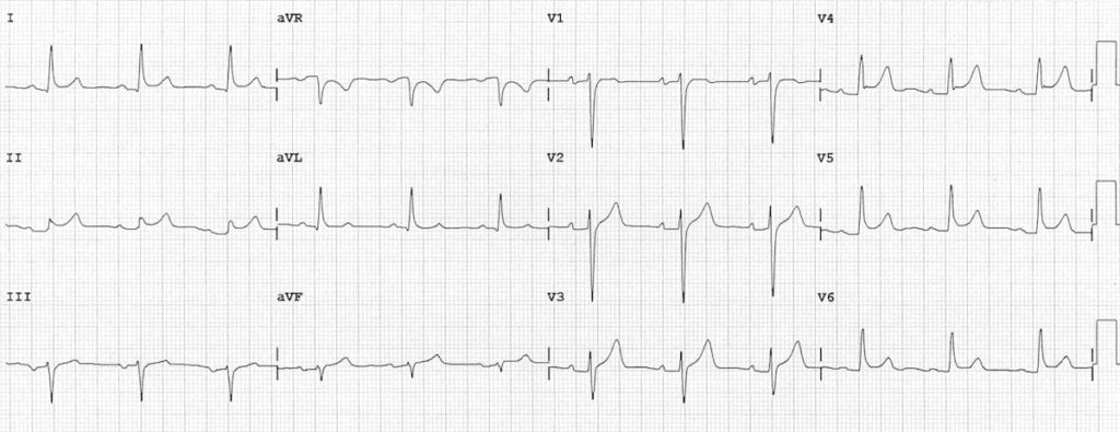
- Early phase is characterized by ST segment elevation, due to inflammation of the epicardium, which accompanies inflammation of the overlying pericardium
- Can have generalized ST-T changes in both anterior and inferior leads
Hyperkalemia:
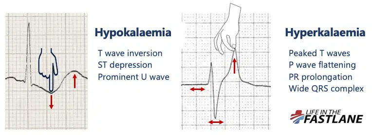
- Distinctive sequence of ECG changes affecting both depolarization (QRS) and repolarization (ST-T)
- First change: Narrowing and peaking of T-waves ('tented' or 'pinched' shape) and can become tall
Hyperkalemia:
- Further elevation: PR intervals become prolonged, P-waves may disappear. Will have intra-ventricular conduction delay, with widening of QRS complexes.
- Can lead to large, undulating (sine wave) pattern with asystole and cardiac death
Inferior Wall Infarction:
- Diaphragmatic portion of the LV
- Will see changes in leads II, III, and aVF
- May produce abnormal Q-waves in these leads
- Generally caused by occlusion of the RCA; less commonly can occur with a left circumflex coronary obstruction
Posterior Infarction:
- Occurs on the posterior (back) surface of the LV
- May be difficult to diagnose because characteristic abnormal ST elevations may no appear in any of the 12 conventional leads
- Tall R-waves and ST depressions can occur in V1 and V2
Q-wave:
- Can occur in any lead; indicates that the electrical voltages are directed away from that particular lead
- With a transmural infarction, necrosis of heart muscle occurs in a localized area of the ventricle
- New Q-waves usually appear within first day of MI
Anterior Wall MI:
- Can see loss of normal R-wave progression in the chest leads (normally should have a progression of height of R-waves from V1-V6)
- In antero-septal infarct, will lose small r waves in V1-V2 (septal depolarization from left to right) and have QS in V1-V2
Myocardial Ischemia:
- One of the most important things to evaluate on EKG
- If severe narrowing/complete blockage of a coronary artery causes blood flow to become adequate, ischemia of the heart muscle develops
- Can be transient (angina pectoris) or more severe (necrosis & MI)
Myocardial Ischemia
- LV consists of an outer layer (epicardium/sub-epicardium) and inner layer (sub-endocardium)
- Can have limit of ischemia to the inner layer or can affect the entire thickness of the ventricular wall (transmural ischemia)
Fascicular Blocks:
- Left bundle branch system: sub-divided into an anterior & posterior fascicle.
- Hemi-block does not widen the QRS complex markedly (compared to a RBBB or LBBB)
Left Anterior Fascicular Block (LAFB):
- Diagnosed by finding of a left axis deviation (-45 degrees or more negative)
- Delayed activation of more superior & leftward position of the LV
- Isolated finding is non-specific; can be seen w/ HTN, AV disease, CAD, and aging