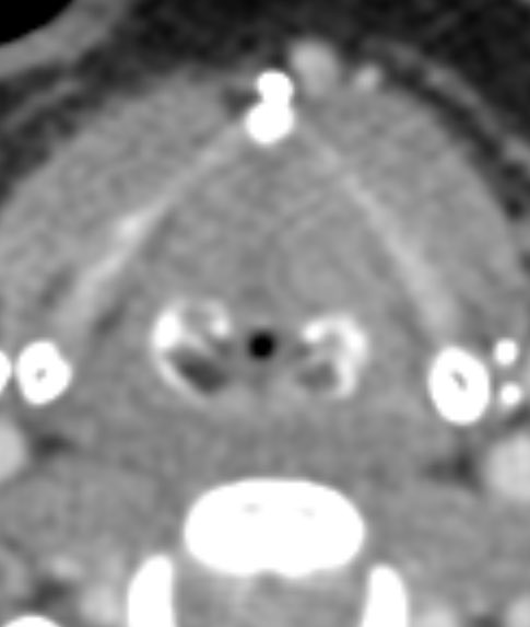
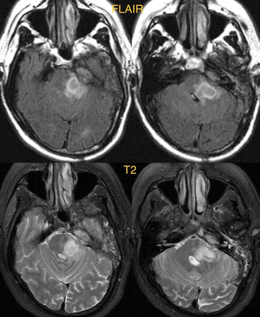
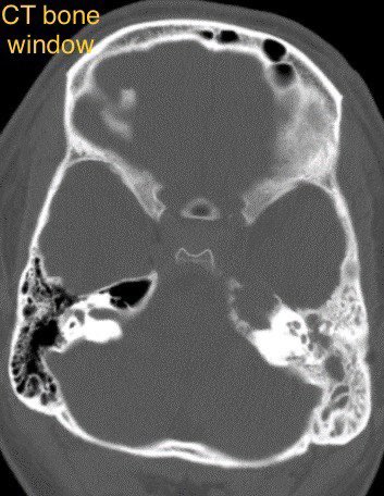
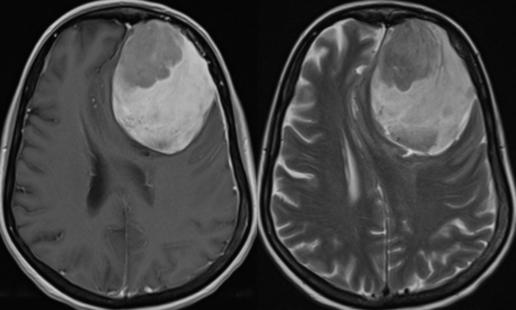
Brain radiation is a risk factor for the development of meningiomas …this patient subsequently developed multiple meningiomas including this large frontal meningioma
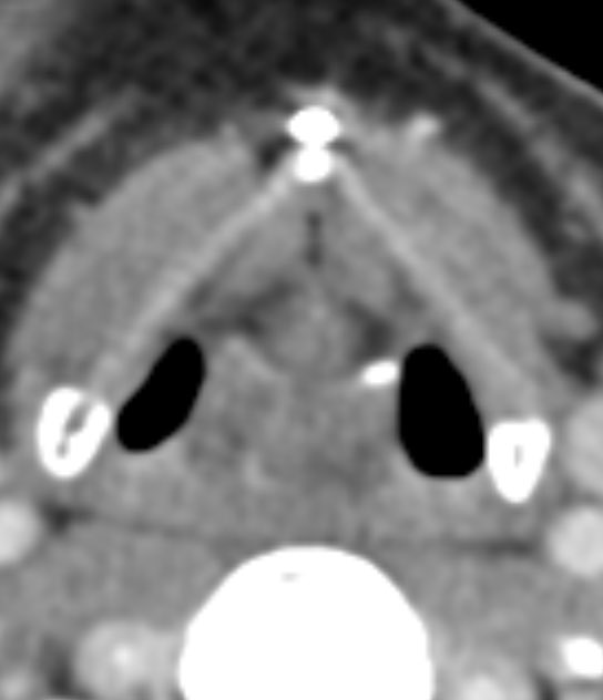
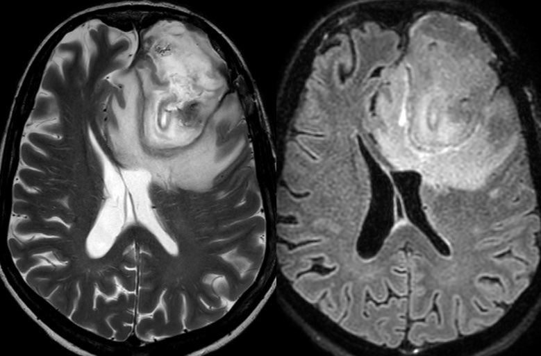
The mass was treated and immediate post op CT shows expected post op change with the resection cavity, some hemorrhage, and pneumocephalus
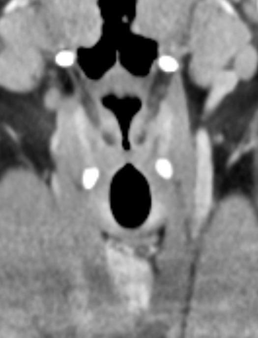
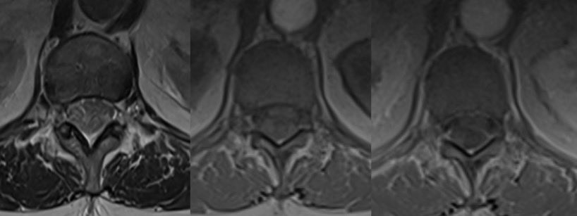
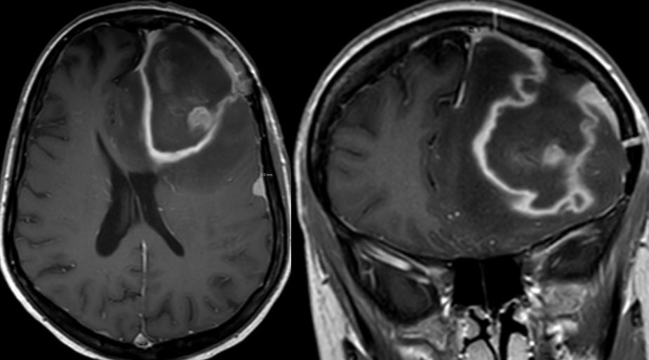
1 month later the patient presents with worsening headaches so an MRI was ordered to eval for post op infection
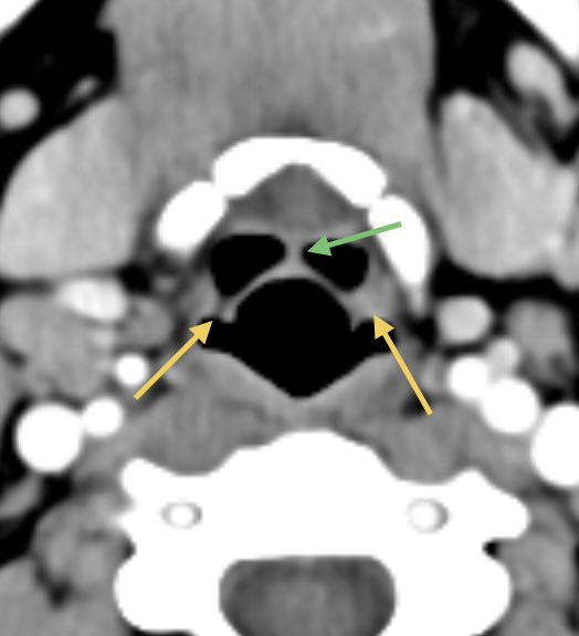
It can be tricky teasing out organizing hematoma from purulent material on MR. CT may not be as helpful as hematoma will reduce in density overtime
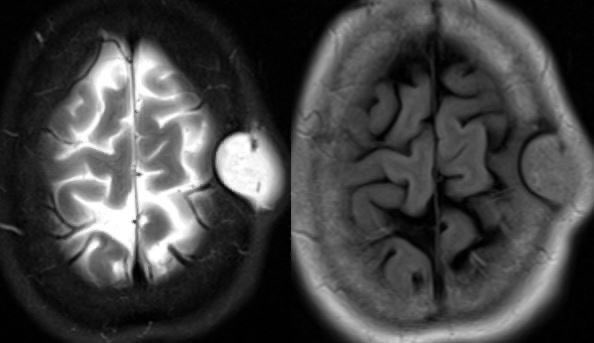
we see here there are areas of restricted and intermediate diffusivity within the resection cavity which can be from purulence or blood product
Given the patchy intrinsic T1 hyperintensity, we know at least some of this is related to subacute blood
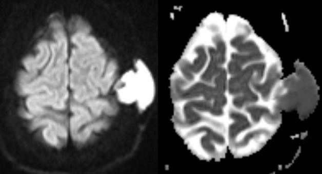
Patchy hypointensity on SWI is also out of proportion to what one would expect from free radical formation along the periphery in abscess, so this is mostly from blood and perhaps foci of pneumocephalus
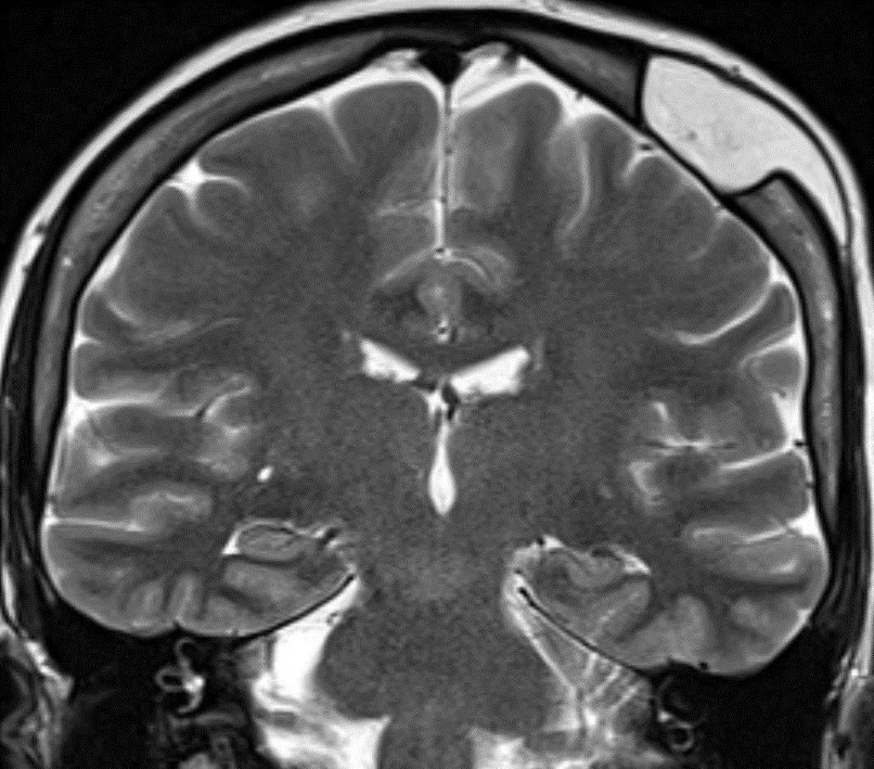
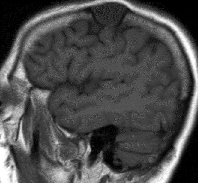
However, there is too much vasogenic edema and mass effect for a 1 month post op. Post op edema and blood should progressively decrease while an infected cavity would increase
OR confirmed both blood and pus
Learning points:
💡 CLINICAL SIGNS OF INFECTION ARE MOST IMPORTANT
💡 PROGRESSIVE or OUT of proportion vasogenic edema suggests infection (though do NOT forget about possibility of tumor recurrence!)
• • •
Missing some Tweet in this thread? You can try to
force a refresh
⭐️ Answer: petrous apicitis complicated by brainstem abscess
🔷Petrous apicitis can display the clinical triad of Gradenigo’s syndrome
1️⃣Otorrhea
2️⃣Cranial nerve 6 palsy
3️⃣Pain in distribution of trigeminal nerve
▶️Petrous apicitis usually occurs as a complication of otomastoiditis when the infection spreads to the skull base.
▶️The petrous apex is in close proximity to Dorello’s canal (where cranial nerve 6 runs through) and Meckel’s cave (where the trigeminal ganglion is located)
▶️CVT causes retrograde venous pressure leading to focal vasogenic edema
▶️Increased back pressure is characterized by dilated veins and petechial hemorrhage which can progress to large hematomas and ischemic neurological damage
🔷Two types of edema can develop:
1️⃣Vasogenic (from venous back pressure)
2️⃣Cytotoxic (ischemia)
🔷Risk factors:
💡 Up to 20% are idiopathic
1️⃣Trauma
2️⃣Tumor/malignancy (compression/invasion from meningioma)
3️⃣Infection
4️⃣Hormonal (pregnancy)
5️⃣Dehydration
🔷What is the most likely diagnosis in this 70 y/o F who lives with feral cats presenting w/ vomiting, diarrhea, leukocytosis, fever for 3 days and progressive decline in level of consciousness?
🔷CSF: initially normal, repeat a few days later ⬆️ WBC (lymphocyte predominant), ⬆️ Protein, normal glucose
🔷 South Central Asia and Southeast Asia appear to have the greatest number of cases
🔷Dogs are the most common reservoir 🐶 (bats in developed countries🦇)
🔷CLINICAL:
▶️Incubation period typically 3 weeks to 2 months (range 5 days to 6 months)
▶️Prodromal symptoms: fever, malaise, anxiety,
and itching at the inoculation site
💡Once in the body, the virus begins retrograde flow to extend to the dorsal root ganglion, which may correlate with neuropathic pain
▶️CNS manifestations: Mental status changes of excessive agitation and depression with hydrophobia and aerophobia
🔷35 y/o F w/ history of Li Fraumeni syndrome presents w/ intermittent left sided weakness and pain. The feeling is of heaviness, difficulty w/ grip, dropping objects and frequent falls. Episodes last for weeks to months. What is your diagnosis? 🧠
🎉Congrats to all the rad fellow matches today!!! 🍾
⭐️ Answer: Tumefactive demyelination (MS in this case)
🔷Imaging in this case is specific enough to diagnosis with confidence but we need extra caution in patients with genetic predisposition to malignancy
💡 In cases where imaging or clinical picture are atypical or discordant for demyelination (especially if there is a genetic disorder), it is best to refer to neurology for proper work up, possible trial of steroids and short imaging follow up
🚩 Be suspicious against MS if your patient has:
1️⃣Systemic symptoms (fever, weight loss, joint or skin symptoms, etc.)
2️⃣Seizures, hearing loss, meningitis signs, movement disorder, aphasia
3️⃣”Family history”
4️⃣Age (<20 or >50)
5️⃣Lesions are symmetric
6️⃣Hemorrhage or dense on CT
7️⃣Diffusion restriction other than leading edge
8️⃣Strokes
9️⃣Cysts
🔟Cortical infiltration
⭐️ 30 y/o presents w/ R sided weakness & fall. Pt had a recent admission ~4 months ago for headache, AMS, seizure & diplopia. Clinically stable until now.