Eduardo R Argaiz Profile picture
Internist | Nephrology Fellow @incmnszmx | Board Member at the Mexican Society of Echocardiography - Academic Committee @SONECOM_AC | #Hemodynamics #EchoFirst
Dr. Jhonny Enrique G Profile picture Julio C. Sauza-Sosa Profile picture paco sarmiento Profile picture 老衲穿白大褂 Profile picture 5 subscribed
Sep 17, 2023 8 tweets 3 min read
👩♀️ Past Medical History: SLE, Antiphospholipid syndrome, portal vein trombosis, ESKD on HD, 🫀Group 1 PH + Severe TR

Now with worsening ascites (Para: SAAG > 1.1, total protein 2.5 g/dL). Lowering dry weigh was attempted..

BP 90/60. No edema. On room air, ⬆️ JVP

#POCUS

1/8 Is this cardiac ascites? Should we lower dry weight even further?

2/8
Aug 4, 2023 6 tweets 4 min read
Hemodynamic Evaluation of Right-Sided Congestion With Doppler Ultrasonography in Pulmonary Hypertension @AmJCardio


50 days' free access link: https://t.co/ADD3F7NgEf

🧵of our findings 👇 (1/6) https://t.co/ORDsb9Nu4rdoi.org/10.1016/j.amjc…
authors.elsevier.com/a/1hXCqgQkyqNA

Image
Image
1⃣ Intra-Renal Doppler (IRVD) alterations are usually classified using morphological patterns (Continuous, Biphasic, Monophasic)

Looking at the relationship between Portal Vein Flow and IRVD you can notice the "Biphasic" pattern shows a very large spread of values! (2/6)
Image
Image
Jul 7, 2023 11 tweets 4 min read
👴 w Cirrhosis ➡️🏥 with spontaneous bacterial peritonitis and septic shock

After fluid resuscitation, vasopressors and antibiotics shock resolved

However now with oliguria and ⬆️ Na (165 meq/L). Cr 1.0 mg/dl, BUN 30 mg/dl

1/10 BP is 155/63 (MAP 94), HR 77, O2 is 94 on O2 8 L/min.

🧠 Encephalopahy on tx w lactulose, edema +++, CRT 1 second, mild ascites.

#POCUS LV/RV OK, LVOT VTI 40 (CO 9.8 L/min), B-Lines, VExUS = 2 (Plethoric IVC + Biphasic Intra-renal Doppler) ➡️ High Output Heart Failure

2/10
Feb 27, 2023 12 tweets 7 min read
HV Doppler from a pt with severe group 1 pulmonary hypertension 👇

Many of us don't have ECG when doing POCUS...

Is it posible to determine this waveform components?

The answer is yes! I'll show you how I did it here

A 🧵on HV Doppler in Pulmonary Hypertension

#VExUS 1/12 Image Normal HV is a mirror image of normal CVP waveform.

It usually has 4 waves:

2 antegrade (flow from liver to 🫀) waves (S and D)

2 retrograde (flow from 🫀 to liver) waves (A and V)

2/12 ImageImage
Jan 29, 2023 13 tweets 7 min read
Young ♀️ w CKD on HD, seen in Cardiorenal clinic

Pt had torrential tricuspid regurgitation due to CVC induced leaflet perforation ➡️ She underwent tricuspid valve replacement surgery 🫀🔪

However, 1 month after discharge she is still using a wheelchair 🤔

1/12🧵 #POCUS above shows plethroic, non-collapsible IVC and Hepatic Veins

Did surgery work?

Is there residual tricuspid regurgitation?

#Echofist (PLAx RV view + A4ch) color Doppler lets us see there is no or minimal TR

Prosthetic valve seems to be working

2/12
Dec 28, 2022 9 tweets 5 min read
Ambulatory Hemodialysis Unit Rounds:

Called to see a patient with hypotension: BP 76/40,🧠 OK, CRT 5 seconds

1st step ➡️🛑Ultrafiltration + 300 ml bolus. BP 90/60

Pt is a middle aged ♂️ w ESRD and T2DM

1/9 🧵 Now 3 kg above Dry Weight.

UF Volume so far: Only 600 ml

🔎📁 Previos HD sessions with no hypotensive episodes

1 week with URI symptoms, 2 days with dyspnea on exertion

On exam: No leg edema, Clear 🫁, JVP hard to assess (hx of multiple CVCs and central vein stenosis)

2/9
Nov 22, 2022 12 tweets 7 min read
#AKIConsultSeries:👨w T2DM➡️🏥 for fever, dysuria and CVA tenderness. On arrival: ⬇️BP, ⬆️Glucose, ⬆️AGMA. Dx UTI + DKA. Tx: Abx + Insulin Pump + 4 L Crystalloid + NE

After resus, pt still oliguric, Cr 3.2. NE 0.7 ug/kg/min,🧠confused, BP 85/62, HR 123, 2L O2. CRT 4 sec

1/12 
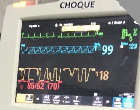
Given DKA, giving additional fluids is tempting. But before we do this, its easy to do a quick assessment of fluid tolerance #POCUS

#LUS shows some B-lines (bilat)
#IVC plethoric w no respiratory collapse
#VExUS shows very pulsatile portal vein 🚨🤔

2/12
Oct 14, 2022 8 tweets 4 min read
🧵Complex #Hemodynamics in ESRD:

Middle age pt ➡️ 🏥 for syncope

HPI: Low BP during HD sessions. Does not achieve dry weight. Today she has had 6 episodes of syncope!

Last episode happened as she stood up from a chair

BP 88/62, HR 87 🧠 OK, CRT 2 sec

1/8 On #Echofist you notice this👇

During systole, flow from the LV towards the Aorta should all be blue in color! (Direction away from the probe)

This apparent change in direction (red sphere) is called Aliasing

Aliasing = Very high flow Velocity!

2/8 Image
Sep 17, 2022 4 tweets 4 min read
Pt seen in ambulatory clinic with worsening kidney function

While the patient is sitting down (90 degrees), you notice neck pulsations!

Are they arterial or venous??

1/4 🧵 It is single peak (but not sharp)

The most striking feature is the inward movement

The breath of movement is diffuse

These are signs of venous pulsations!

Very helpful table from @AndreMansoor 👇


2/4
Aug 20, 2022 13 tweets 8 min read
Young pt ➡️ 🏥 worsening shortness of breath

PMH: ESRD. Only 1 HD session/week. However, residual urine volume has now decreased substantially

On exam: BP 134/94, 2L O2,🧠✅, elevated JVP, decreased 🫁 sounds at bases, No murmurs, very mild edema. Functional left BC AVF

1/13 Careful examination of neck veins reveals no pulsations, even with pt sitting up 🤔

What could explain the absence of venous pulse? 2/13
Jul 10, 2022 11 tweets 7 min read
#AKIConsultSeries Middle-aged male ➡️🏥 for painful knee and fever. Now in shock 🚨

📂Chart review: PMH EtOH Cirrhosis, right knee arthroplasty.

It is always a good practice review previous PACS images🩻: Nodular liver, colateral vessels and prosthetic right knee

1/11 On exam: BP 72/48, HR 82, O2Sat 95%.
CRT 7 sec, 🧠somnolent, confused. No edema, no obvious ascites.

Warm, swollen and erythematous knee: Tap with obvious purulent fluid🧫

Cr 2.8 mg/dl (baseline 0.5), K 6.7, Urine 🔬: hyaline casts, some urothelial cells

2/11
Mar 17, 2022 10 tweets 4 min read
#AKIConsultSeries 👵➡️🏥 w necrotizing fasciitis s/p debridement. Significant 🩸 during surgery ➡️ intensive resuscitation (transfusions + crystalloid)

48 hrs later 💧📈 =+13 L, Edema +++, Hypoxemia, pleural effusion.

Now anuric unresponsive to high dose IV diuretics 1/10 Image 
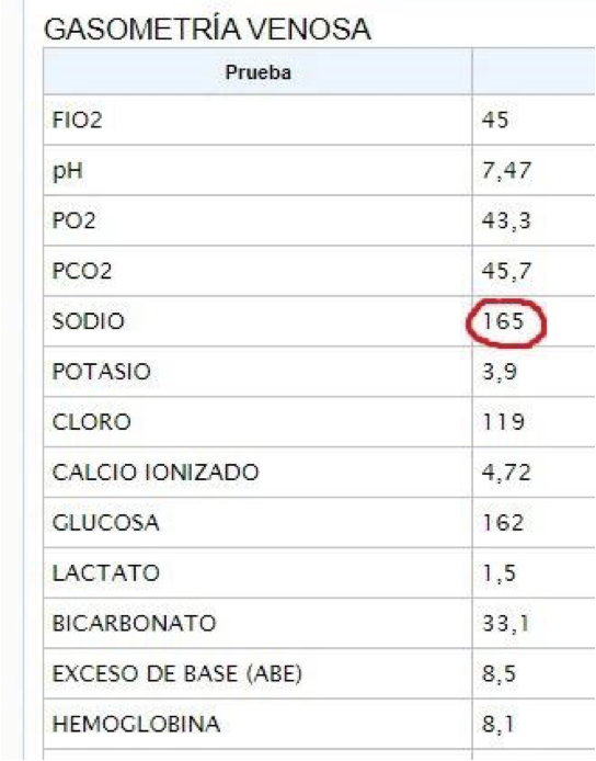
Cr 3.2, K 3.5, HCO3 25, Hb 8.9, WBC 26k, 95% PMN, Lactate 2.5

MAP 65 on NE (0.02 ucg/kg/min), HR 130, O2 sat 80-85 on 15 L NRB, RR 17 but w increased respiratory effort.🧠 drowsy, CRT >8 sec👇, JVP not elevated

Primary team consults for RRT initiation with ultrafiltration 2/10
Dec 23, 2021 10 tweets 4 min read
Patient with flank pain, hematuria and significant leg edema

#POCUS 🧵

1/10 Lung Ultrasound #LUS 2/10
Dec 8, 2021 13 tweets 7 min read
A tale of two hearts: Physiological observations on AV shunts and congestion 🧵

These are 2 patients on IHD I saw in the outpatient clinic

🔷 Both with severe venous congestion (#VExUS = 3)
🔷 Both with tortuous brachiocephalic AV fístula

1/11 What I found remarkable was the diametrically opposed effects of manual AVF compression on JVP! 🤯

🔴 Patient A: AVF Compression improves venous congestion
🔵 Patient B: AVF Compression worsens venous congestion

2/11
Nov 28, 2021 5 tweets 4 min read
Pt with advanced Cirrhosis

AKI, Oliguria + Encephalopathy

Initial treatment = Albumin + Lactulose enemas

🧂Na is now 158 (From Lactulose induced free water loss)

#POCUS ninja @tumleal noticed something was wrong! He got his probe and texted me

📱 #WhatsAppAKIConsult 1/5 #
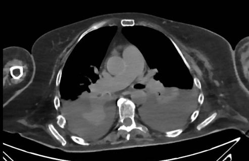
POCUS: Plethoric non collapsible IVC

Based on the clinical scenario, IVC, heart rate and Pulse Pressure...

What is the likely underlying pathophysiology? 2/5
Oct 21, 2021 6 tweets 3 min read
Pt w right HF and high probability pulmonary hypertension

TAPSE 15 mm, TRVmax 4.1 m/s, paradoxical septal motion

Renal Venous Doppler 👇

According to doi.org/10.1161/JAHA.1…, Which curve color would best describe this patient's PH-related morbidity?

Poll and 🧵👇

1/6 ImageImage Which curve in the Kaplan Meier Curve above best fits this particular patient?

2/6
Aug 28, 2021 18 tweets 9 min read
One of my favorite and most intriguing causes of severe venous congestion (#VExUS = 3)

A 🧵on High Output Heart Failure (HOHF) 1/18 First, the index case:

Clip above shows hyperdynamic flow from the vena cava

#echofirst 👇: Very dilated and plethoric IVC, LV OK, Dilated RV, D sign

Overall: Increased Right heart filling pressures

2/18
Jul 13, 2021 7 tweets 4 min read
#AKIConsultSeries

👴 w Advanced Cirrhosis, hemorrhagic portal hypertension and hx of a heart block (w pacemaker).

Came to the ED w diarrhea 2/2 severe C. Diff.

Now in shock...

Initial resus with Norepinephrine 0.3 ug/kg/min, Crystalloid and albumin. Cr 3.8

1/7
18 hrs later, no renal improvement + oliguria.

MABP 70, CRT 2 seconds. 🧠 confused, + asterixis. No ascites or edema

Workup: hemodynamic AKI (⬆️SG, ⬇️UNa, ⬆️BUN/Cr, bland sediment)

Team wants to continue fluids, albumin and antibiotics... Dr. Harris, do you concur?

2/7
May 3, 2021 12 tweets 7 min read
AKI Consult: 👵 ➡️ ED with severe DKA. CT Abdomen and Chest to look for infectious trigger: negative. Tx with IV insulin and balanced crystalloid + 6 L with obvious improvement. Cr was 2.7

Remained oliguric, now in sudden shock with increasing NE dose (0.5 ucg/kg/min) 🚨 1/12 #POCUS Very hyper-dynamic🫀 with increased contractility and no RV dysfunction.

🔎 Look carefully at color of flow exiting the LV:

Aliasing (green color): This means ultrasound system is trying to image an event that is occurring faster than the sample rate

2/12
Feb 16, 2021 11 tweets 6 min read
Pt w advanced liver cirrhosis. 🏥 Comes w worsening ascites. No fever🤒, no bleeding🩸. 🧠 ok, no asterixis. BP 91/50. Labs📈: AKI (Cr 3.0 mg/dl), UNa 7 mEq/L, bland sediment. #POCUS 👉small cirrhotic liver with significant ascites. Paracentesis ruled out PBE. 📊Poll below👇 1/11 What would your initial treatment be? 2/11
Feb 10, 2021 15 tweets 7 min read
📞 Nurse: Patient has a blood pressure of 226/118 mmHg!
📞 Resident: Nifedipine 30 mg STAT!
.....
📞 Me: About that last call, please hold Nifedipine until we assess the patient

A 🧵of some cases of Inpatient Hypertension 👇 1/15 🔎🖥️..This was a pt w ARDS on IMV. Other vitals 🆗. Previous BP= normal, no recent change in sedation, vent 🆗, no asynchronies. UOP = 0 for 2 hrs 🤔. Exam: Distended bladder!

After foley catheter change, BP normalised 😎

Why do we have this reflex to treat acute high BP? 2/15