Galway University Hospitals Dept of Anaesthesiology & ICM -Online educational resource for Anaesthesia, ICM, advanced critical care echo and clinical research
Apr 5, 2022 • 9 tweets • 3 min read
1/9 Tuesdays Tweetorial:
You are in cardiothoracic theatre doing a mitral valve replacement for severe MR
The anaesthetic consultant is doing a TOE and keeps saying 'PISA'
U know its in Italy but have no idea why he keeps saying it
What is PISA and why is it used?
2/9 PISA = Proximal Isovelocity Surface Area
Blood is ejected -> LA
It converges at the mitral regurgitant orifice it forming hemispheres -> different blood velocity in each hemisphere
RCCs that are equidistant from the orifice(in each hemisphere) travel at similar speed
Mar 15, 2022 • 7 tweets • 2 min read
Quiz answer:
1.Sir Austin Bradford Hill
2.The Bradford Hill Criteria – 1965
Guidelines to help assess whether an observed association between an exposure and an outcome is likely to be causal.
If known severe MR – these are the NEW findings that should prompt you to speak to you friendly cardiologist colleagues
Sep 14, 2021 • 14 tweets • 7 min read
1/13 - GUH Echo tweetorial:
The tricuspid regurgitation jet velocity shown was used in a critical care patient to estimate RV systolic pressure:
Vol control – tvol 420ml PEEP 10cmH20
Cardiovascular: MAP 67mmHg on Noradrenaline 0.3mcg/kg/min
2/13
His TRVmax is high:
Why should I not diagnose this patient with pulmonary hypertension in my echo report?
2 are correct:
a. not steady state
b. Off axis cursor
c. Echo cannot diagnose it
d. Poor 2D view
Sep 14, 2021 • 4 tweets • 6 min read
GUH Images in Anaesthesia and ICU:
The tricuspid regurgitation jet velocity shown was used in a critical care patient to estimate RV systolic pressure:
Vol control – tvol 420ml PEEP 10cmH20
Cardiovascular: MAP 67mmHg on Noradrenaline 0.3mcg/kg/min
His TR Vmax suggests his RV systolic pressure is 51mmHg + RA pressure = HIGH
I am conscious that I should not diagnose this patient with pulmonary hypertension in my echo report:
Why?
Jun 24, 2021 • 15 tweets • 6 min read
1/14
GUH - Echo Tweetorial - Aortic Stenosis:
How do they calculate: 1. Valve area 2. Mean AV gradient 3. Max AV gradient
2/14
Essential Principles: 1. The effective orifice area is always smaller than the anatomical orifice area
This effective orifice area is what is calculated
It is the key determinant of survival 2. Continuity equation
Conservation of mass
Jun 22, 2021 • 11 tweets • 7 min read
GUH - images in Anaesthesia and ICU:
In anticipation of our Level 2 echo tweetorial series
This is a zoomed image of a valve during a level 2 scan
1. Name the valve
Name the valve:
Mar 21, 2021 • 6 tweets • 3 min read
1/6
Answer:
LA myxoma
And for those of you who still auscultate the precordium you would have heard the elusive 'tumour plop'....of course you would.
But lets get back to the basics of M- Mode use in the PLAX view
2/6
Firstly the AV:
Here is a normal m-mode image through the AV during the cardiac cycle - note: 1. How systole and diastole are identified by ECG 2. Opening of the RCC and NCC to form the 'envelope' 3. Symmetry of the envelope 4. Closure line at end syst.
Feb 1, 2021 • 10 tweets • 5 min read
1/10
Echo Tweetorial 4 - SUBCOSTAL VIEW
1. The subcostal view 2. IVC 3. Additional subcostal views Incredibly versatile TTE view especially in ventilated patients but is trickier than it seems But first - Hand movements!
2/10
So what are we looking at:
Nov 26, 2020 • 15 tweets • 9 min read
1 Welcome to #researchmethodologies with @DrAoifeBee
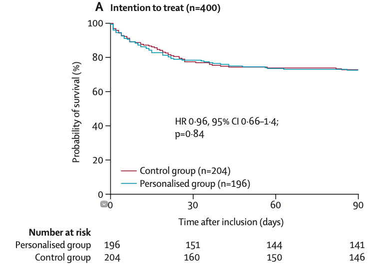
Kaplan-Meier (KM) curves are a wonderfully informative way of presenting survival outcomes over time. But how do we interpret them? Survival analysis determines the probability of a binary outcome (aka an event or a failure)
2/15
Survival means the event being studied has not occurred yet - the patient is still alive if you’re analysing mortality, the baby has not been delivered if analysing births, the patient has not yet met whatever criteria you have decided constitutes an event in your study.