
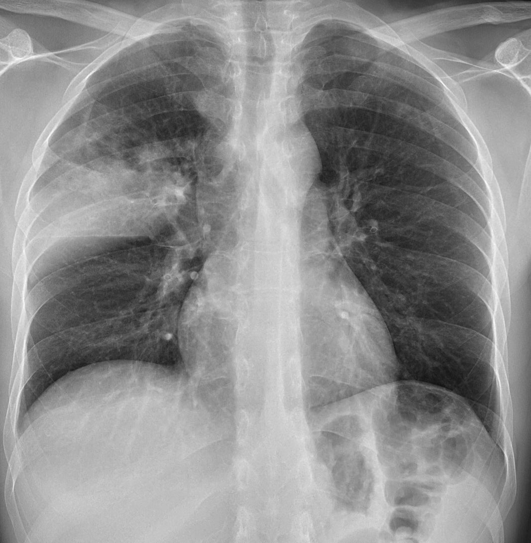
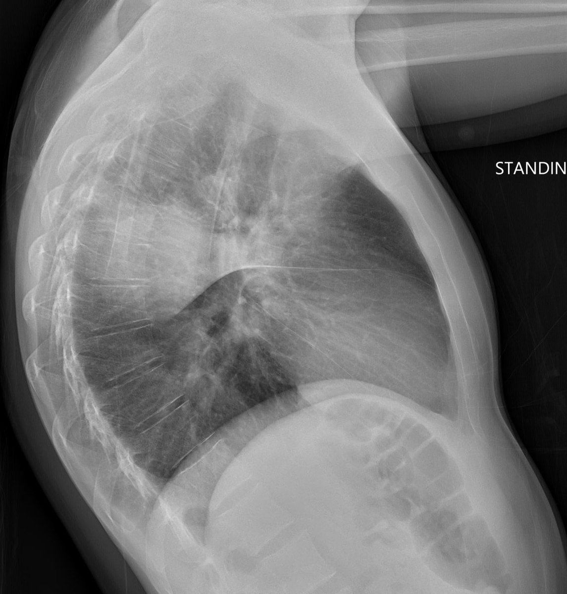
 This is classic lobar pneumonia, involving the right upper lobe. Sometimes pna can look mass like on one of the views, but often the other view shows it spreading out along a fissure or other pleural boundary, fading along the non pleural margin, more typical of pna than cancer.
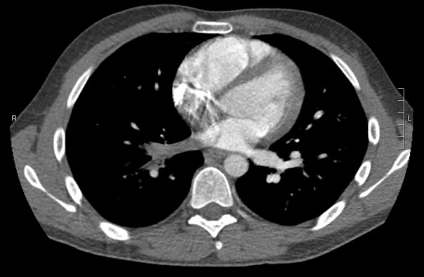 Here are the lung windows, showing pretty typical right lower lobe pneumonia. Does this change your thinking about the first image? Note that there is no interstitial edema in the RLL or pleural effusion.
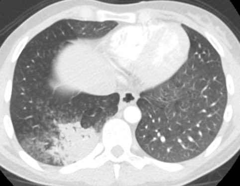
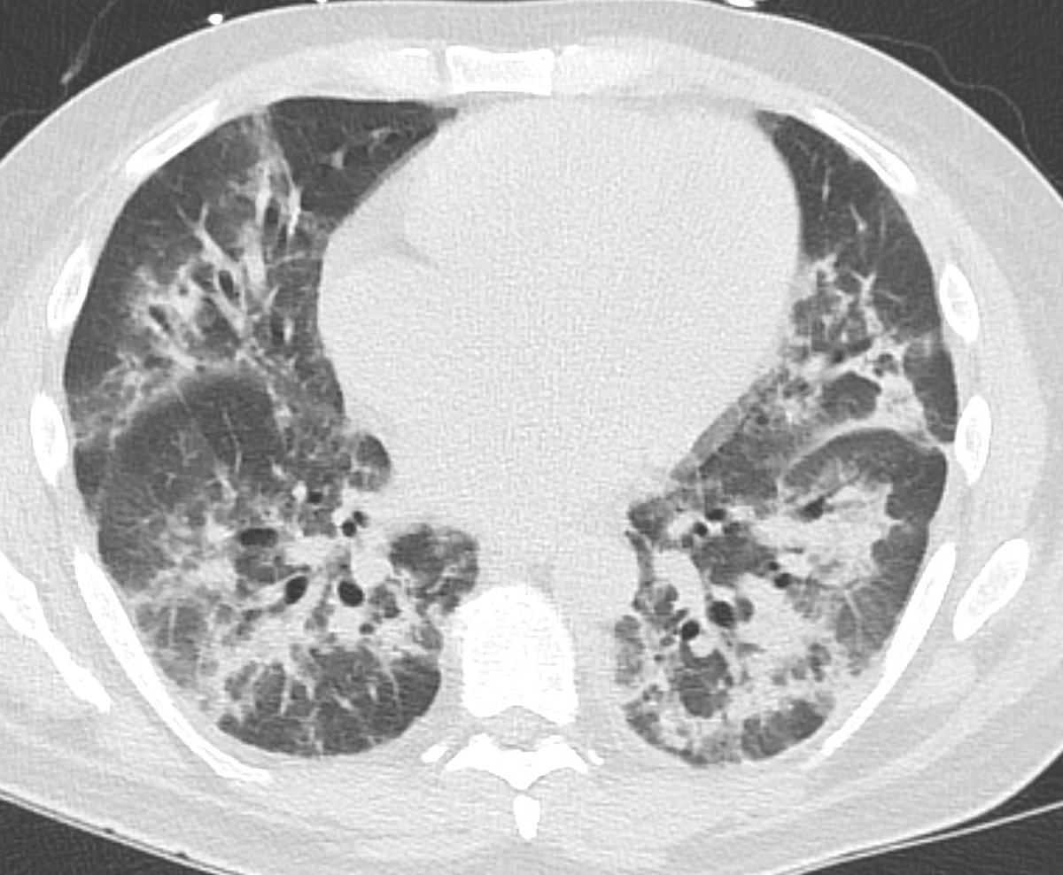 The patient is a 45 yo male. Acute kidney injury and ? Pneumonia. The first image was at presentation. There is peribronchial consolidation with a nice perilobular appearance peripherally. Looks like organising pna. Cause could be infection, vaping, drug reaction, CTD...