Marcus Pinto, MD, MS Profile picture
Peripheral Nerve Neurologist and Pathologist @MayoClinic. Check my Highlights for Neuropathy Education. Special Interest in Amyloidosis, Vasculitis and Trials
Mar 28 7 tweets 4 min read
Good morning, #NeuroX; I hope you all are having a great week in the clinic and will not feel down learning that Dr. Peter Dyck could identify the etiology of 76% of the “idiopathic” neuropathies referred to him in the 1970s.

In this post, I break down one of my favorite papers of all time.

This is the second post in celebration of Dr. Dyck’s retirement. I will try to post two more next month to compensate for February (sorry, busy times here 😅).

1/x

onlinelibrary.wiley.com/doi/abs/10.100…Image
Image
These were patients seen by Dr. Dyck and/or Dr. Ed Lambert only. Eighty percent of these patients were referred by neurologists. Each patient underwent a detailed history and examination, along with nerve conduction studies (NCS), electromyography (EMG), quantitative sensory testing (QST), cerebrospinal fluid (CSF) analysis, and, in some cases, nerve biopsy.

If the neuropathy phenotype indicated a potential inherited neuropathy, family members of the patients who might also have neuropathy were invited to the clinic for neurologic evaluations, EMG, QST, and, in certain cases, nerve biopsy. If no family members had a history of neuropathy, parents, siblings, and children were invited to the clinic for the same evaluations.

2/x
Mar 12 10 tweets 6 min read
The lumbosacral plexus plays a vital role in motor, sensory, and autonomic innervation of the lower limbs and pelvic region.

Lumbosacral plexopathies are not easy to diagnose, and below, I try to simplify how to approach them.

The main focus of this post is clinical features and diagnosis, but we will also touch on anatomy and treatment.

The temporal profile (TEMPO) is critical when assessing lumbosacral plexopathies. We divide them based on the time from symptom onset to nadir in hyperacute (< 24 hours), acute/subacute (1-30 days), and chronic (> 1 month).  The hyperacute causes are usually traumatic or vascular, which could be ischemic or compressive, secondary to retroperitoneal hematoma.  The acute/subacute causes are usually inflammatory (lumbosacral radiculoplexus neuropathies-LRPN) or infectious (syphilis, CMV, or VZV).  The chronic etiologies are usually inflammatory, radiation-induced, or neoplastic.

The most common causes of lumbosacral plexopathies I see in my practice are LRPN and radiation-induced plexopathies. Given my very specialized clinic, I also see very rare causes, like paraneoplastic, focal CIDP, and neurolymphomatosis, so I approach all of them the same way.

1/xImage
Image
The lumbosacral plexus originates from the ventral rami of the L1-S4 nerve roots and consists of two main sections: the upper lumbar plexus and the lower lumbosacral plexus. The upper section is formed by the L1-L4 nerve roots, occasionally with input from the T12 root. Meanwhile, the lower section is derived from the L4-S4 nerve roots. Like the brachial plexus, structural variations, such as prefixed or postfixed configurations, are often observed. Anatomically, the plexus is embedded within the psoas major muscle and emerges from its lateral border. It provides motor and sensory innervation to the lower limb and pelvic region on the same side.

2/x
Jan 31 10 tweets 2 min read
When I have a new fellow or resident in our peripheral nerve clinic, I always teach them the importance of defining the neuropathy clinical syndrome. NCS/EMG are valuable, but the neurological exam is the most important (as it should be in neurology but many have forgotten).

This is the neuropathy clinical phenotype classification I find the most helpful:

- Isolated small fiber neuropathy
- Length-dependent peripheral neuropathy (or distal symmetric polyneuropathy)
- Multiple mononeuropathies
- Mononeuropathy
- Asymmetric neuropathy
- Polyradiculoneuropathy
- Plexopathy (brachial or lumbosacral, many times a radiculoplexus neuropathy)
- Sensory neuronopathy
- Motor neuronopathy

I always tell my trainees they don’t have to memorize the causes of each phenotype. If you define the syndrome correctly, you can ask Google or ChatGtp what are the most likely causes. 😊

Definitions 👇👇👇

#NeuropathyBites

1/x
Isolated Small Fiber Neuropathy

Pure small fiber neuropathy. Only temperature and pinprick dysfunction on exam. Ankle reflexes must be normal in patients < 60 yo. NCS must be normal for age.

2/x
Jan 15 8 tweets 3 min read
Many of my neurology colleagues feel uneasy when admitting a patient with severe, chronic (> 2 months) rapidly progressive peripheral neuropathy of uncertain etiology. While there are more than 100 causes of neuropathy, the most likely causes in this clinical setting can be narrowed down to the six below:

🔺 Chronic Inflammatory Demyelinating Polyradiculoneuropathy (CIDP), including autoimmune nodopathies

🔺 Amyloidosis

🔺 Paraneoplastic syndrome, including POEMS syndrome

🔺 Vasculitis, including radiculoplexus neuropathies

🔺 Lymphoma or carcinomatous meningitis

🔺 Nutritional deficiencies

#NeuropathyBites
#NeuroTwitter
#MedTwitter
#MedX

1/x
It’s important to note that this post refers to patients with moderate to severe disability caused by neuropathy, often dependent on wheelchairs and doesn't include patients with multifactorial gait disorder. In addition, some electromyographers report “severe neuropathy” based solely on lower extremities conduction studies, so caution is advised when using nerve conductions to define the severity of a neuropathy.

Another critical point is that patients are not good at differentiating progression versus no improvement of subacute/chronic weakness, so use landmarks like starting gait aids, the ability to stand up from the toilet without using the hands or help of someone, and falls, foot drop, wrist drop, abnormal gait, or inability to do something in their daily routine when they started. Be mindful that GBS, by definition, does not progress beyond 4 weeks.

Amyotrophic Lateral Sclerosis (ALS) is a more common cause of hospital admissions than all the conditions listed above, so a thorough history and detailed examination of the patient are essential. Bulbar symptoms/signs and upper motor neuron signs are highly suggestive of ALS. Moreover, up to 20% of ALS patients may also exhibit concomitant peripheral neuropathy; therefore, be careful about “motor-predominant polyradiculoneuropathies” that present with only mild sensory involvement.

2/x
Jan 9 11 tweets 3 min read
In celebration of the legendary career of Professor Peter J. Dyck, who officially retired last month, I kick off today this series with a hidden gem: The 10 P's in the characterization and differential diagnosis of peripheral neuropathy.

#NeuropathyClassics

#DyckPapers

1/ Image
Image
Modern medical educators may not agree with a 10 point mnemonic but this was the 80s/90s and Dr. Dyck is a very thoughtful and comprehensive neurologist.

neurology.org/doi/10.1212/WN…

The first “P” (P1) is for Pattern:

- Anatomical and temporal localization of the disease.

- Anatomical pattern includes mononeuropathy, monoradiculopathy, multiple mononeuropathies, plexopathy, polyneuropathy or polyradiculoneuropathy.

- Temporal pattern considers onset (acute, subacute, chronic) and course (monophasic, recurrent,
progressive).

2/
Dec 30, 2024 5 tweets 1 min read
Acute nutritional axonal neuropathy (dry beri-beri) is more common than many believe.

In patients with malnutrition, intractable vomiting, heavy alcohol use, or a poorly balanced diet, this condition is more likely than Guillain-Barré Syndrome (GBS). #NeuropathyBites

1/
These patients need IVT(thiamine) and not IVIg. I treat them with 500 mg IV q8h for 3 days, then 100 mg daily orally, and supplement other vitamins as needed.

The neuropathy is usually more distal and sensory predominant compared to GBS, though it can mimic GBS exactly.

2/
Dec 2, 2024 7 tweets 2 min read
🚨Check out our latest work on vasculitic myopathy @GreenJournal! A fantastic collaboration between @MayoClinicNeuro and #MayoRheum. Thank you, @ElieNaddaf3, for the incredible mentorship, and @MdWarrington and #MattKoster for the help. Breakdown ⬇️ 1/7
neurology.org/doi/pdf/10.121… • We report 25 patients vasculitic myopathy

• Primary systemic Vasculitis: 40% (ANCA vasculitides most common)

• Secondary systemic vasculitis: 48% (RA most common)

• Nonsystemic vasculitic myopathy: 12%

2/7 Image
May 20, 2020 11 tweets 7 min read
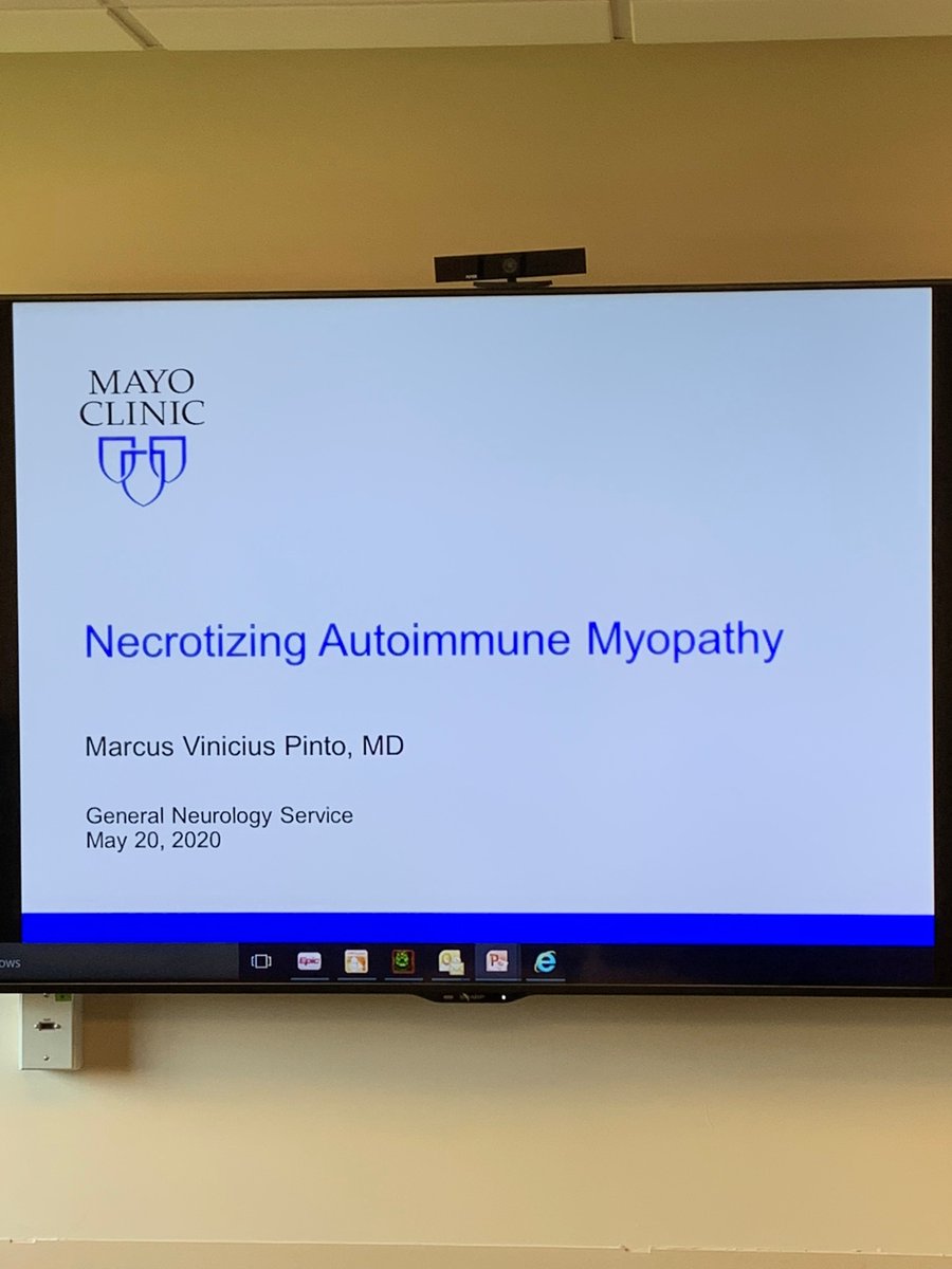
Feels so good to be back to #Neurology. Today I had the pleasure to give #Neurologymorningreport on Necrotizing Autoimmune Myopathy #NAM. #MedTwitter follow below a few pearls I shared with the team @ErsidaBuraniqi @nzalewski2. THREAD. #NAM is an autoimmune myopathy characterized by severe proximal weakness, myofiber necrosis with minimal or no inflammatory infiltrates on muscle biopsy, and infrequent extra-muscular involvement.