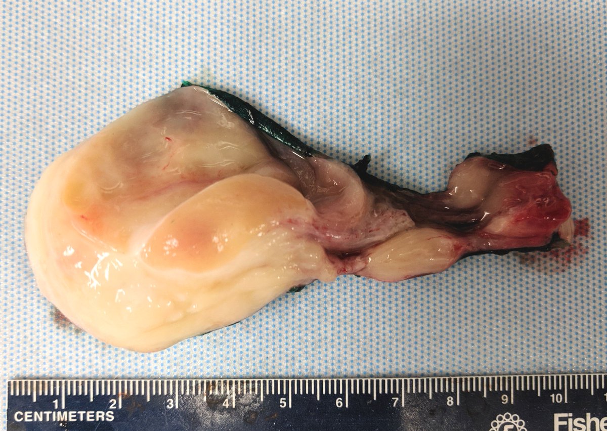


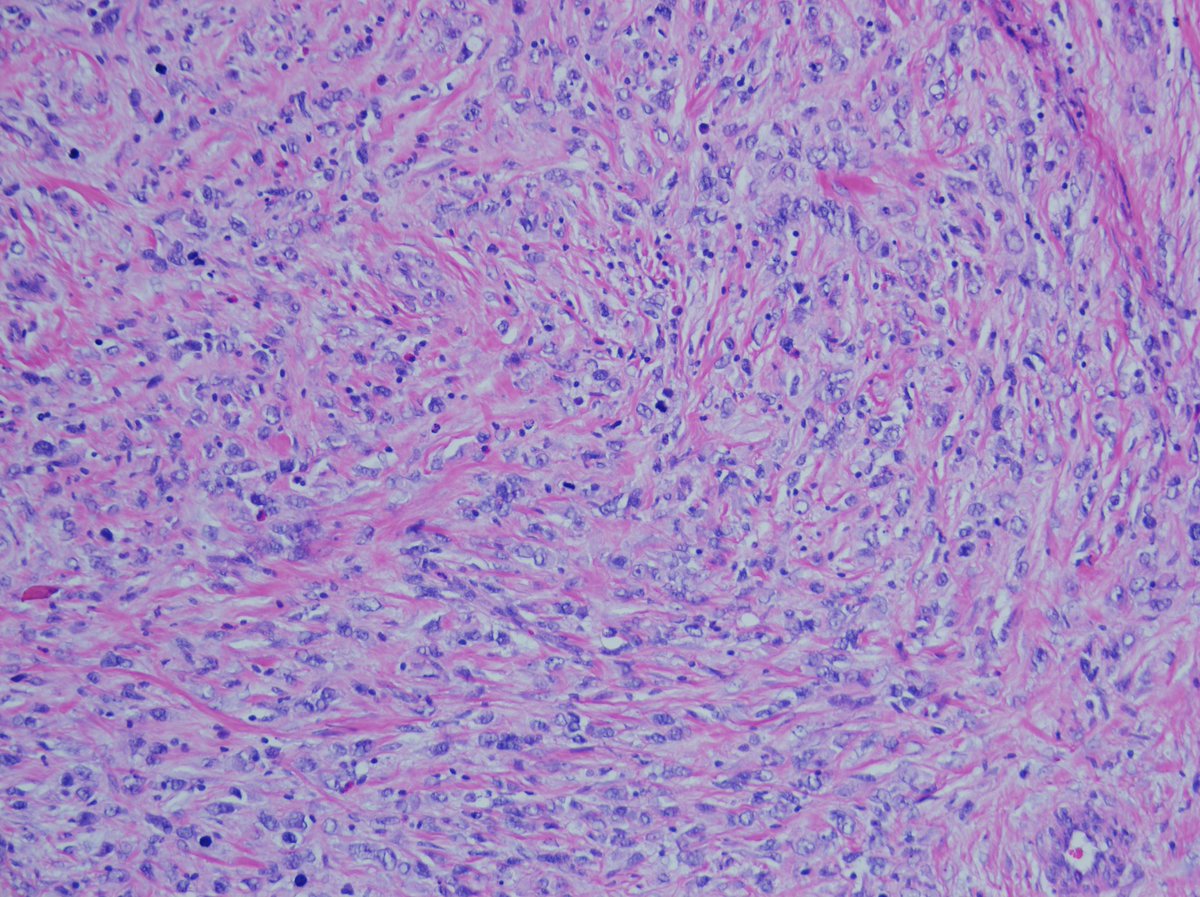
Rhabdomyosarcoma is indeed the top-level diagnosis... but more importantly, what type??
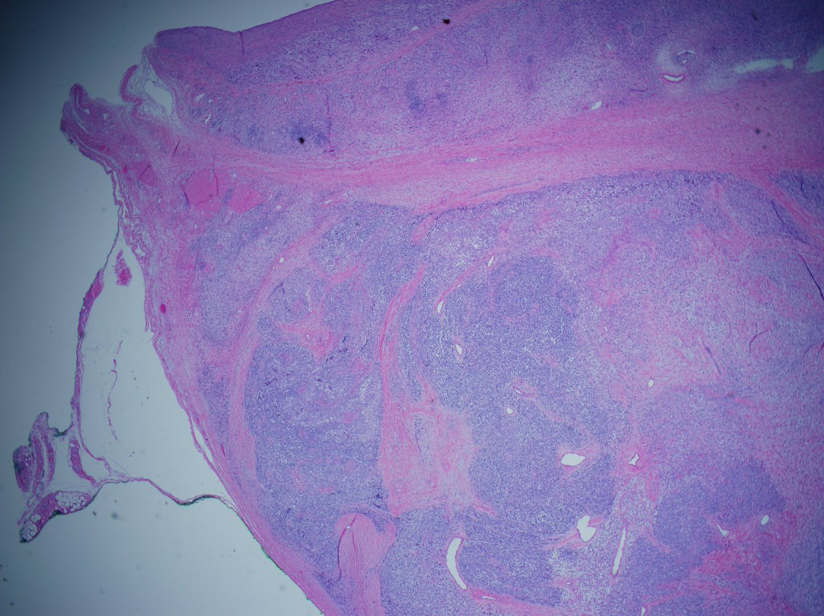
Note: several H&E patterns here that correspond nicely to the patterns on cytology.
#pedipath #gupath #pathology
Note: several H&E patterns here that correspond nicely to the patterns on cytology.
#pedipath #gupath #pathology
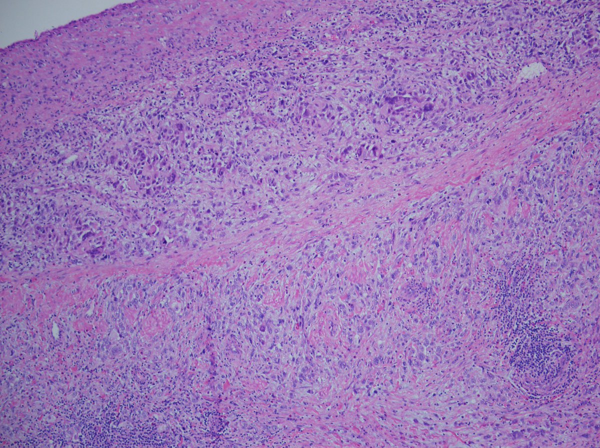
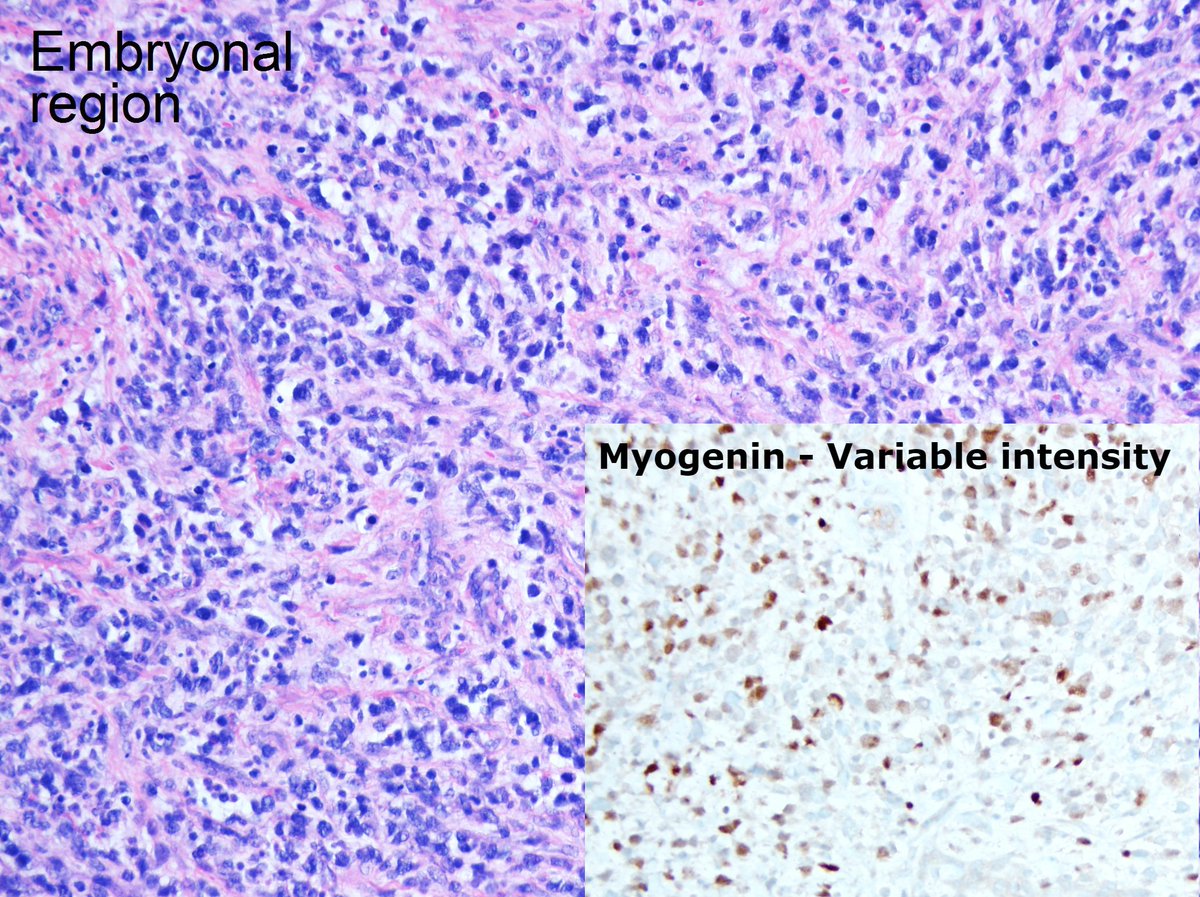
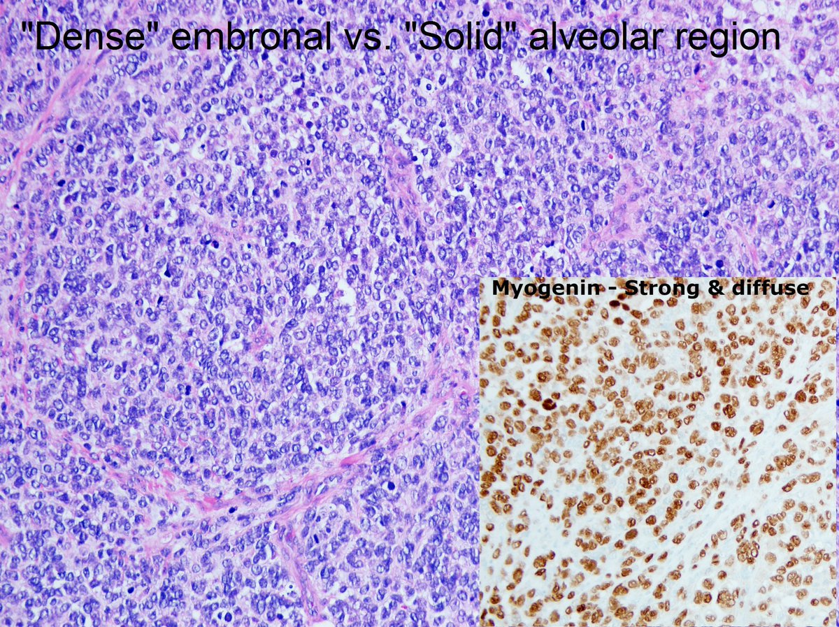
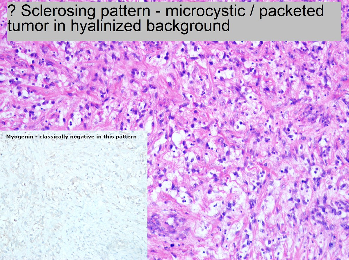
Note the multiple patterns here - and see how they relate to myogenin staining.
This is an uncommon example of mixed-histology rhabdomyosarcoma.
Classic Embryonal - variable myogenin
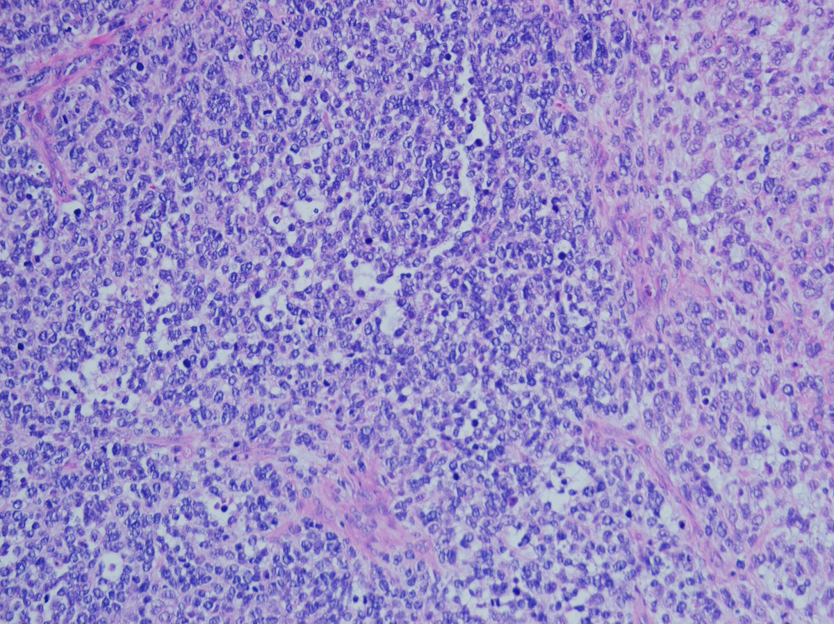
Dense Emb/Solid Alveolar - strong, diffuse myogenin
Sclerosing - Weak/absent myogenin
#pedipath
This is an uncommon example of mixed-histology rhabdomyosarcoma.
Classic Embryonal - variable myogenin
Dense Emb/Solid Alveolar - strong, diffuse myogenin
Sclerosing - Weak/absent myogenin
#pedipath

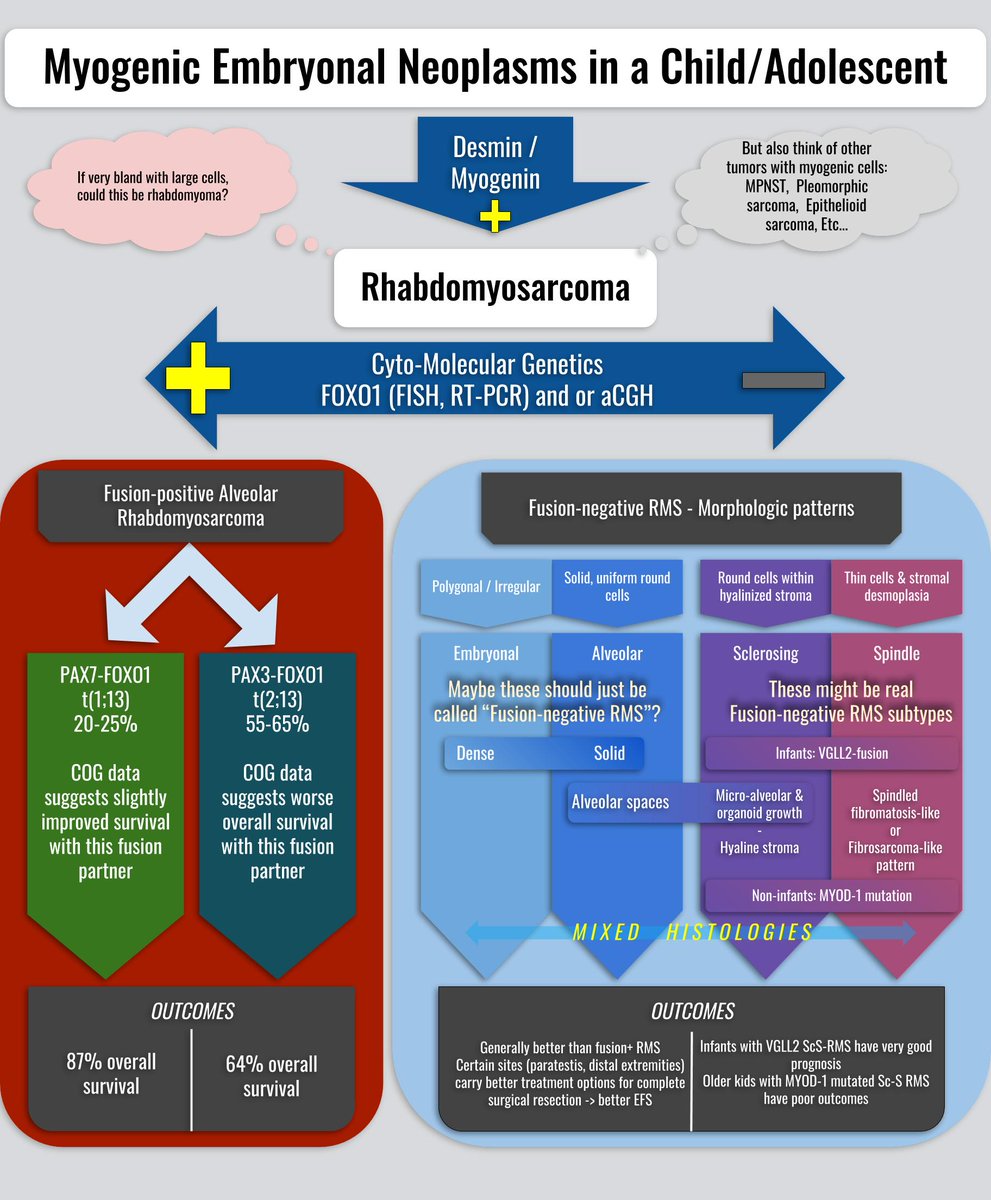
To summarize:
There is genetic evidence that ERMS and fusion-neg ARMS are the same thing. May be that in the near future, RMS will be subdivided as
FP-RMS (PAX7),
FP-RMS (PAX3)
FN-RMS
ScS-RMS (VGLL2)
ScS-RMS (MYOD mut)
Large 👇🏽
drive.google.com/open?id=15-8rZ… …
#pedipath #bstpath
There is genetic evidence that ERMS and fusion-neg ARMS are the same thing. May be that in the near future, RMS will be subdivided as
FP-RMS (PAX7),
FP-RMS (PAX3)
FN-RMS
ScS-RMS (VGLL2)
ScS-RMS (MYOD mut)
Large 👇🏽
drive.google.com/open?id=15-8rZ… …
#pedipath #bstpath
Forgot to close the loop on this case.
Given varied histology and presence of "solid alveolar" vs "dense embryonal" histology, we performed FISH, PCR and array cGH on two separate block.
ALL methods failed to show a FOXO1 fusion.
Thus, this is: fusion-negative ERMS
Given varied histology and presence of "solid alveolar" vs "dense embryonal" histology, we performed FISH, PCR and array cGH on two separate block.
ALL methods failed to show a FOXO1 fusion.
Thus, this is: fusion-negative ERMS