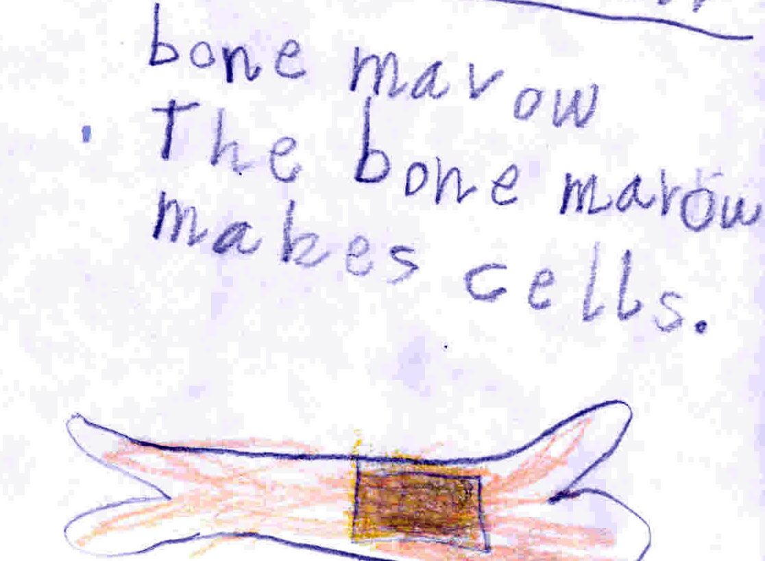
#HematologyTweetstory 18: bone marrow aspiration & trephine biopsy - critical diagnostic tests in evaluating suspected hematologic disease. Here’s a diagram of the procedure for patients (from @MayoClinic) & illustration of the basic concept from my then 6-year-old daughter.🙂/1 

Two things surprise me about the history of marrow biopsy. First: marrow sampling only quite recently became part of routine hematology practice, even though the importance of marrow morphology in diagnosing disease was recognized in the mid-1800s. (Images: @ASH_hematology) /2

Second: it took a long time to transition from sternal to iliac crest aspirates. Surely someone leaning over a patient to do a sternal aspirate circa 1945, worrying about a fatal mediastinal incursion like the ones below, thought, “There must be a better way to do this”? /3


Humans have long known how to get inside bone. Prof. Tracy Kivell @UniKentSAC posits that eating marrow was a key historical driver of our species survival. Marrow is the most calorie-rich food left after apex predators abandon a kill, and fueled our big energy-hungry brains/.4


Then for centuries, bone was penetrated via trephining. This medical practice was common in pre-Colonial Mesoamerica & the Andes. My friend @vmontori took me to a museum in Peru with numerous trephined skulls. Healing around wounds suggests many patients survived the procedure./5


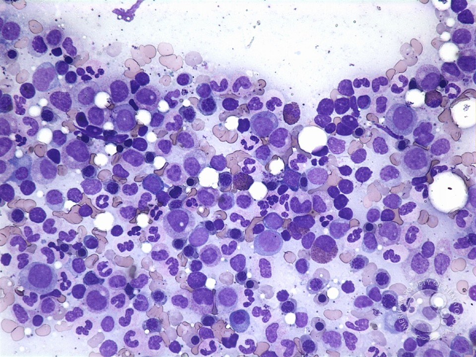
In the 19th century it became clear that marrow was where blood cells originated. Impressively, Ernst Neumann worked that out in the 1860s without histologic stains, which weren't invented until 1870s. LL is a picture of what unstained marrow looks like. Hard to see anything!/6


So all 19th century observations about disease pathology were taken from marrow samples obtained post-mortem. For example, in 1846 John Dalrymple described a patient with "mollities ossium" (now called #myeloma); this detailed drawing of plasma cells was obtained after death./7


In 1903, Alfred Wolff-Eisner (1877-1948) in Berlin trephined the tibia and femur of several animals and suggested bone marrow biopsy during life might be useful clinically. He was also one of many people who observed large "lymphoid" cells in the marrow - we call them blasts./8


In 1905, Giuseppe Pianese (1864–1933) in Naples (statue) reported marrow sampling from femurs of living people. He used a small curette, similar to "marrow spoons" once popular for eating marrow. His goal was not to assess marrow histology per se, but to look for leishmaniasis./9


Similarly, in 1908 Giovanni Ghedini of Genoa reported accessing the tibia of some living patients - he was also searching for microorganisms - with trephining tools. That didn't really catch on, which is good since the tibia is usually hypocellular in adults & non-diagnostic./10


Then Leipziger Carly Seyfarth (1890-1950) used trephining tools in 1923 for evaluating sternal marrow in cases of suspected malaria. Unfortunately this approach was traumatic and caused a lot of bleeding and infections, so didn’t catch on./11


I found out Seyfarth went to Russia in 1922-23 as part of a humanitarian Red Cross expedition. He spent time working & teaching in this former "Alexander Hospital" in St. Petersburg. I think this tweetorial may be the first time that visit has been linked to what happened next/12

In 1927, Mikhael Arinkin (Михаил Иннокентьевич Аринкин; 1876-1948) at a nearby military hospital in Leningrad (where Ivan Pavlov, of dog salivation fame, also worked) modified Seyfarth’s technique: he used a thin lumbar puncture needle for sternal aspiration. Much safer! /13


Other's modified Arinkin's technique by introducing special needles with guards to prevent entering too deeply, pioneered by M. J. Arjeff in Germany 1931 and Rudolf Klima in Vienna in 1935. One image is from a great 2007 @BritSocHaem review by L. Parapia of Bradford UK./14


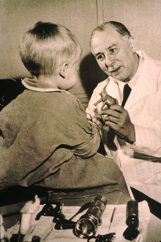
A couple of Belgians, L. van den Berghe and I. Blitstein, were the first to use iliac crest to obtain marrow in 1945. But it didn’t catch on! In 1949 for example, William Dameshek compared sites: yes that really is a needle in a spinous process of a child, briefly in vogue./15

In 1950 Michael A. Rubinstein and Amiel Smelin @MontefioreNYC said "anterior iliac crest > sternum" as aspirate site; in 1952, Howard Bierman @cityofhope suggested *posterior* iliac crest would be even better, which is how most marrows are done today (as below diagrams show)./16


Another interesting marrow event happened in 1952. A 4 y.o. girl in Baltimore, Ann Theresa O’Neill, was diagnosed with ALL & treated @SaintAgnesMD. She developed measles & was near death. As she was dying, a ward Sister prayed for intercession from Elizabeth Seton (1774-1821)./17


Against all odds, the girl got better, as this 1994 @washingtonpost article describes in detail (with quote from Ron McCaffrey @BrighamHeme ). /18
washingtonpost.com/archive/lifest…
washingtonpost.com/archive/lifest…
Today we would attribute her complete response to Coley’s toxins brought on by the measles, but the Sisters were convinced it was a miracle - and at some level maybe both are true. In 1961, Ann was examined by Sidney Farber @DanaFarber and Joe Burchenal (R) @sloan_kettering./19

Ann underwent many sternal and iliac marrows; she was in remission. In 1962, based on this & 2 other miracles, Seton was beatified. In 1975 she became 1st American-born Catholic saint. Dr. Farber laughed at the irony, “It fell to me, a Jew, to prove cure so she could be sainted.”

I used to see patients each week @StElizabethsMC in Brighton (a Boston neighborhood) -hospital named after a different St Elizabeth but w/ several Seton icons. The library there is named after Fred Stohlmann Jr, former editor of @BloodJournal, who came to a tragic end in 1974./21


Stohlmann & his wife were flying home from Tel Aviv #ISH meeting in 1974 when their airplane exploded over the Aegean. Legendary MPN doctor Murray Silverstein was scheduled to be on that flight but had too much to drink and missed it; he later claimed drinking saved his life./21

Back at Mayo, Murray's colleague Malcolm Hargraves, discovered the LE cell - an early test for lupus - by walking between hospitals with a vial of marrow in his shirt pocket. Hargraves was an early environmental activist & gave Rachel Carson some data for “Silent Spring”./23


In 1971, Khosrow Jamshidi, an Iranian physician, patented a special needle with a cutting edge and a tapered interior. This is still the most commonly used marrow biopsy today. His patent diagram is below. Today these needles often come in pre-packaged kits./24


A brief pause in this tweetstory to consider how yummy bone marrow can be, and what it looks like uncooked. Consider this a snack break as we head into the home stretch. @Bloodman /25


@Bloodman In some places sternal aspirates are still routine. But not here in the US anymore. There are rare circumstances in which sternal aspirates are still done here, eg those patients with fat distributions such that no conventional needle is long enough to reach the iliac crest./26
I performed sternal aspirate on this man for diagnosis after negative iliac crest biopsies; a PET scan showed uptake in the sternum. Wonderful haematologist @CKBrierley from @MRC_WIMM Oxford who was visiting us at the time wrote up that case for #AmericanJournalofHematology. /27

Sternal aspirates are still risky, and I worry every time even though I'm incredibly careful and have now done dozens. (They’re far safer than bedside spleen biopsies, which used to be routine diagnostic procedures. As you can imagine, complication rate from that was high.)/28

A new innovation is use of automated drills to enter the marrow space. We've done some work with OnControl system; its quick & the core samples are often larger than manual Jamshidi. (I have no COI.) However we don't often use it, since more costly. Others do routinely use it./29

Also, some hospitals always do marrow biopsies in radiology with CT guidance. I think the main advantage is that this saves time and effort for hematologists-oncologists who may rarely do marrow aspirates/biopsies. But CT biopsy is so more expensive & rarely truly necessary./30

Once marrow is extracted, it can be prepared for analysis in different ways. But that’s another story, for another day! (These stories take many hours to research & collect images) This image is from Meyerson, Concise Guide to Hematology 2018./31

So here at the end, I hope I'm allowed one editorial comment. 🙂 We're living in a time when international scientific collaborations are under greater scrutiny than before. In recent days, there have been many prominent people dismissed or even arrested for undisclosed ties./32
Some of these situations look really bad, even treasonous. We must to prevent stealing and espionage. But also... this was a story to which Americans, Europeans, Asians, and so many from all over the world of diverse religions & worldviews have contributed./33
I'm not naive; there are folks who are greedy or bad actors. Yet we're all better off if we can still find ways to collaborate as *humans* to move medicine & science forward. We can't make it harder to work across borders because some have abused foreign ties. That's it.😉 /33End

• • •
Missing some Tweet in this thread? You can try to
force a refresh