
1/
You're on call and open a chest CT for a patient with suspected #COVID19.
How do you interpret and report the imaging findings?
A #TWEETORIAL of the @Radiology_RSNA Expert Consensus Statement on Reporting Chest CT Findings Related to COVID-19
pubs.rsna.org/doi/10.1148/ry…
You're on call and open a chest CT for a patient with suspected #COVID19.
How do you interpret and report the imaging findings?
A #TWEETORIAL of the @Radiology_RSNA Expert Consensus Statement on Reporting Chest CT Findings Related to COVID-19
pubs.rsna.org/doi/10.1148/ry…

2/
This consensus statement has been endorsed by @thoracicrad and @RadiologyACR.
Important work by our faculty @PennRadiology Drs. Scott Simpson & Harold Litt.
Podcast: rsnaradiologycti.libsyn.com/welcome-to-the…
*Figures/tables in this #tweetorial from orig. manuscript, unless otherwise stated.
This consensus statement has been endorsed by @thoracicrad and @RadiologyACR.
Important work by our faculty @PennRadiology Drs. Scott Simpson & Harold Litt.
Podcast: rsnaradiologycti.libsyn.com/welcome-to-the…
*Figures/tables in this #tweetorial from orig. manuscript, unless otherwise stated.
3/
🔑KEY POINT:
☝️First, it’s important to note that routine screening chest CT for identification of #COVID19 is NOT currently recommended by most professional organizations.
@CDCgov Guidance:
cdc.gov/coronavirus/20…
@RadiologyACR Recommendations:
acr.org/Advocacy-and-E…
🔑KEY POINT:
☝️First, it’s important to note that routine screening chest CT for identification of #COVID19 is NOT currently recommended by most professional organizations.
@CDCgov Guidance:
cdc.gov/coronavirus/20…
@RadiologyACR Recommendations:
acr.org/Advocacy-and-E…
5/
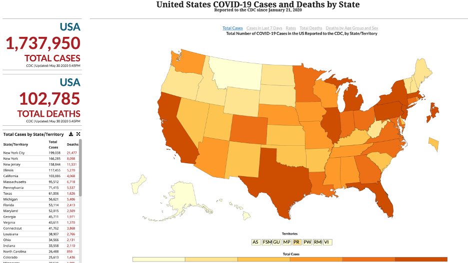
Here’s a look at #COVID19 cases & deaths in the United States by State as of 5/30/2020
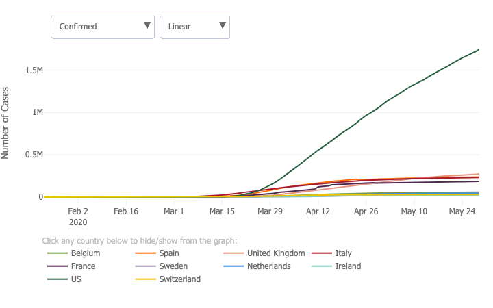
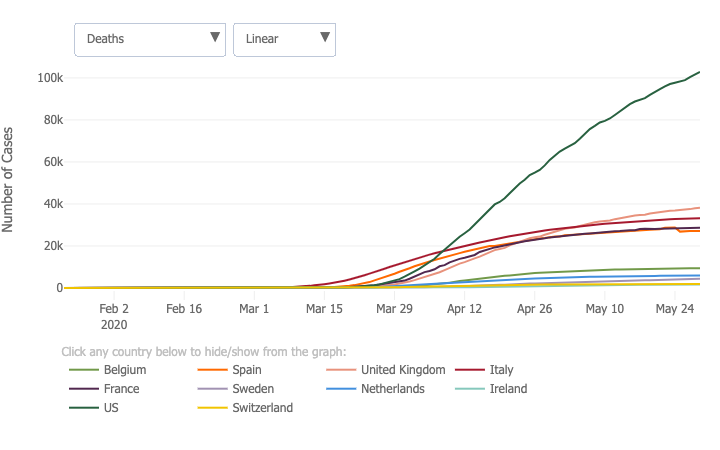
And a look at confirmed cases & deaths for the 10 countries with the highest absolute number of deaths, including the US (dark green).
Figure sources: @CDCgov and @JohnsHopkins
Here’s a look at #COVID19 cases & deaths in the United States by State as of 5/30/2020
And a look at confirmed cases & deaths for the 10 countries with the highest absolute number of deaths, including the US (dark green).
Figure sources: @CDCgov and @JohnsHopkins
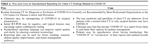
Should you include "COVID-19" in the radiology report of a patient with chest CT findings potentially attributable to #COVID19 pneumonia?
6/
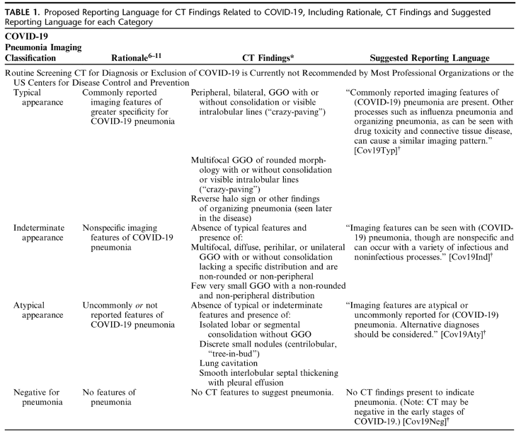
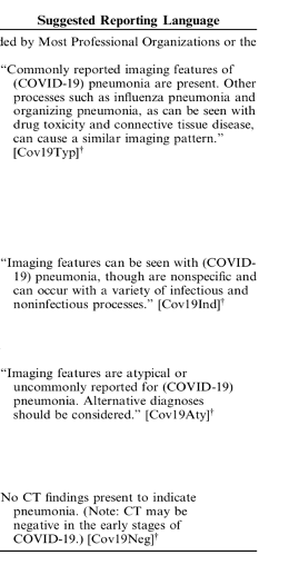
The authors propose 4 categories for reporting CT imaging findings related to #COVID19 with suggested standardized reporting language.
1) Typical appearance
2)Indeterminate appearance
3)Atypical appearance
4)Negative for pneumonia
The authors propose 4 categories for reporting CT imaging findings related to #COVID19 with suggested standardized reporting language.
1) Typical appearance
2)Indeterminate appearance
3)Atypical appearance
4)Negative for pneumonia

10/
🔑KEY POINT:
Chest CT findings can precede positivity on RT-PCR.
🔑KEY POINT:
Chest CT findings can precede positivity on RT-PCR.
12/
What imaging features would be considered #indeterminate for #COVID19?
🔘Non-rounded, non-peripheral GGO
🔘Lack of specific distribution
🔘Can be multifocal, diffuse, perihilar, or unilateral
🔘+/- consolidation
What imaging features would be considered #indeterminate for #COVID19?
🔘Non-rounded, non-peripheral GGO
🔘Lack of specific distribution
🔘Can be multifocal, diffuse, perihilar, or unilateral
🔘+/- consolidation
13/
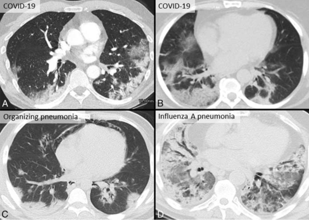
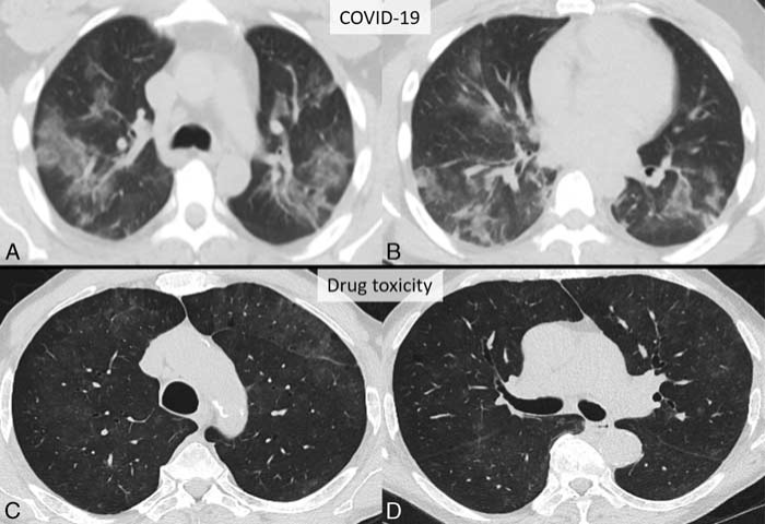
#Indeterminate Cases:
Patchy GGO with non-rounded morphology and without specific distribution in two patients.
🔘(A,B): Patient with #COVID19 pneumonia
🔘(C,D): Patient with acute lung injury from presumed drug toxicity.
#Indeterminate Cases:
Patchy GGO with non-rounded morphology and without specific distribution in two patients.
🔘(A,B): Patient with #COVID19 pneumonia
🔘(C,D): Patient with acute lung injury from presumed drug toxicity.
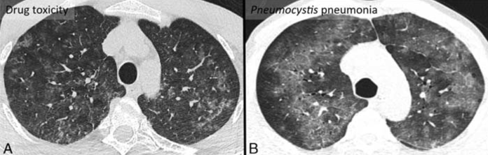
14/
#Indeterminate Cases:
Diffuse GGO without specific distribution & with non-rounded morphology in two different patients.
🔘Patient A: findings were the result of acute lung injury from presumed drug toxicity
🔘Patient B: findings were the result of Pneumocystis pneumonia.
#Indeterminate Cases:
Diffuse GGO without specific distribution & with non-rounded morphology in two different patients.
🔘Patient A: findings were the result of acute lung injury from presumed drug toxicity
🔘Patient B: findings were the result of Pneumocystis pneumonia.

Does a negative chest CT exclude the possibility of #COVID19?
19/
Now, it's time to generate our #radiology report.
How should we report the findings?
The authors:
🔘Make the case for structured reporting
🔘Propose standardized language
🔘Cover the pros and cons of standardized reporting for chest CT findings related to #COVID19.
Now, it's time to generate our #radiology report.
How should we report the findings?
The authors:
🔘Make the case for structured reporting
🔘Propose standardized language
🔘Cover the pros and cons of standardized reporting for chest CT findings related to #COVID19.
21/
The authors recommend direct communication with the referring provider to discuss likelihood of viral infection and reach consensus.
When noted incidentally, findings do not need to be reported as #COVID19 pneumonia, and “viral pneumonia” is a reasonable alternative.
The authors recommend direct communication with the referring provider to discuss likelihood of viral infection and reach consensus.
When noted incidentally, findings do not need to be reported as #COVID19 pneumonia, and “viral pneumonia” is a reasonable alternative.
22/
💥TAKE HOME POINT # 1
The goal of this expert consensus is to help #radiologists recognize & report imaging findings of #COVID19 pneumonia.
But remember, consultation with clinical colleagues at your institution is key to establishing an agreed upon reporting approach.
💥TAKE HOME POINT # 1
The goal of this expert consensus is to help #radiologists recognize & report imaging findings of #COVID19 pneumonia.
But remember, consultation with clinical colleagues at your institution is key to establishing an agreed upon reporting approach.
23/
💥TAKE HOME POINT # 2
Despite most professional guidelines recommending against routine screening CT for #COVID19, chest CTs may be requested for diagnosis & management, particularly when RT-PCR is not readily available.
💥TAKE HOME POINT # 2
Despite most professional guidelines recommending against routine screening CT for #COVID19, chest CTs may be requested for diagnosis & management, particularly when RT-PCR is not readily available.
24/
💥TAKE HOME POINT # 3
Standardized chest CT reporting language can provide a consistent reporting framework, improve clarity and reduce variability when reporting chest CT findings related to #COVID19.
💥TAKE HOME POINT # 3
Standardized chest CT reporting language can provide a consistent reporting framework, improve clarity and reduce variability when reporting chest CT findings related to #COVID19.
@threadreaderapp @covid19threads
unroll
unroll

