Wondering whether axonal input *drives* fMRI-based functional connectivity? Us too!
Here is what we found using #chemogenetic deconstruction of rsfMRI in the mouse🐁🐭
➡️Cortical silencing results in paradoxical fMRI overconnectivity tinyurl.com/yy269j92
Thread below 1/n
Here is what we found using #chemogenetic deconstruction of rsfMRI in the mouse🐁🐭
➡️Cortical silencing results in paradoxical fMRI overconnectivity tinyurl.com/yy269j92
Thread below 1/n
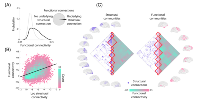
Computational and empirical evidence suggest that structural and functional connectivity are robustly related
➡️ this recent review from @richardfbetzel @misicbata summarizes it all tinyurl.com/yyy5u3zm
2/n
➡️ this recent review from @richardfbetzel @misicbata summarizes it all tinyurl.com/yyy5u3zm
2/n
⚠️A key prediction of structurally based models of fMRI coupling is that *inactivation* of a brain node would result in reduced rsfMRI connectivity with its targets⚠️
But is that really the case?
3/n
But is that really the case?
3/n

➡️Seminal computational modelling supports this hypothesis tinyurl.com/y4rqucmn
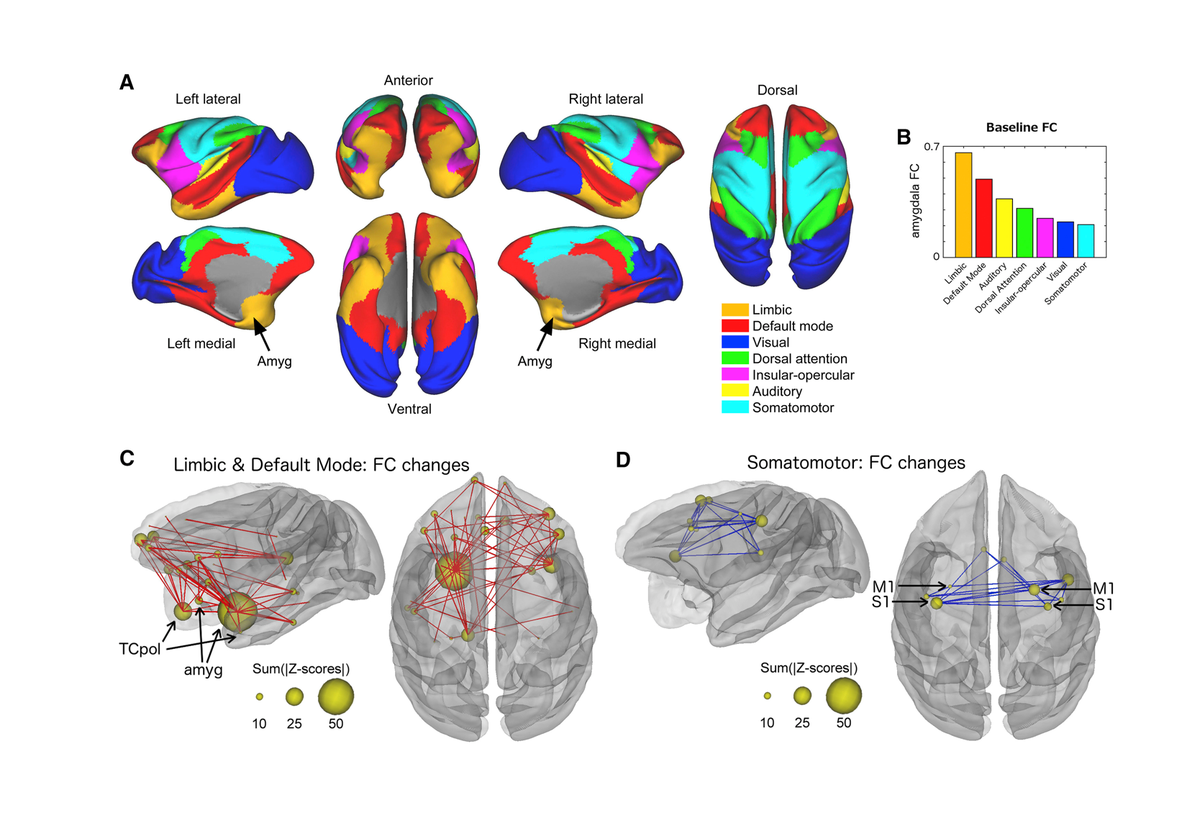
➡️Chemo-fMRI work in primates does too, as inactivation of the amygdala leads to reduce rsfMRI connectivity as predicted
tinyurl.com/y5zv57vf
So this settles the question, right?
4/n
➡️Chemo-fMRI work in primates does too, as inactivation of the amygdala leads to reduce rsfMRI connectivity as predicted
tinyurl.com/y5zv57vf
So this settles the question, right?
4/n
Well, as often the case in science, things might be more complicated than they seem
For example intact bilateral networks have been observed in acallosal people tinyurl.com/yxtwvd7w
5/n
For example intact bilateral networks have been observed in acallosal people tinyurl.com/yxtwvd7w
5/n
Plus *over-connectivity* is often observed in disorders characterized by loss of cortical function Alzheimer's Stroke.. tinyurl.com/y2pnfxuh
So there seems to be also evidence arguing AGAINST a purely dyadic relationship between structural and functional connectivity..
6/n
So there seems to be also evidence arguing AGAINST a purely dyadic relationship between structural and functional connectivity..
6/n
...which led us to investigate how inactivation of a cortical region *causally* affects brain-wide fMRI connectivity
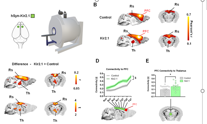
➡️We first chronically silenced the 🐭prefrontal cortex via overexpression of a K+ channel. ePhys below shows that this leads to reduced spontaneous firing
7/n
➡️We first chronically silenced the 🐭prefrontal cortex via overexpression of a K+ channel. ePhys below shows that this leads to reduced spontaneous firing
7/n

Surprisingly, we found that chronic PFC silencing results in *increased* rsfMRI connectivity between the silenced area and its targets within the mouse default mode network (midline thalamus, posterior cingulate - retrosplenial in rodents)
8/n
8/n

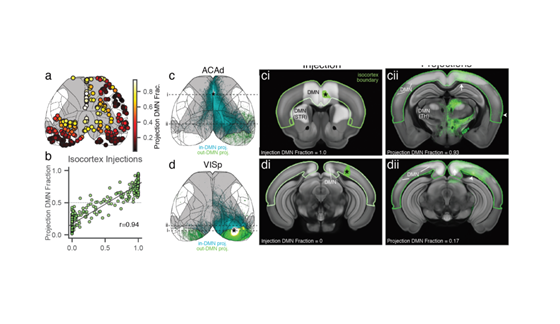
By comparing overconnected regions against a voxel-resolution model of the structural connectome tinyurl.com/y4n5mtl8 we found that all the affected regions are robustly innervated by the PFC
➡️ these are areas that are directly "communicating" with the PFC
9/n
➡️ these are areas that are directly "communicating" with the PFC
9/n

Could this effect reflect homeostatic/neuroadaptive adaptations? What happens if we acutely silence the PFC instead?
To address these questions we designed a chemo-fMRI study in which DREADDs were used to acutely silence the mouse PFC
10/n
To address these questions we designed a chemo-fMRI study in which DREADDs were used to acutely silence the mouse PFC
10/n

We found that acute DREADD inhibition of PFC reproduces the same pattern of rsfMRI *overconnectivity*
This means
➡️effect is not manipulation specific
➡️nor a neuroadaptive response to protracted inhibition
Plus, with DREADDs we can now investigate neural mechanism
11/n
This means
➡️effect is not manipulation specific
➡️nor a neuroadaptive response to protracted inhibition
Plus, with DREADDs we can now investigate neural mechanism
11/n

Using in vivo ePHYS we found that DREADDs robustly and specifically attenuates firing in PFC
This lead to
➡️*reduced* ƴ activity (expected)
➡️*increased* δ power (intriguing!)
Could δ band coupling explain the observed overconnectivity?
12/n
This lead to
➡️*reduced* ƴ activity (expected)
➡️*increased* δ power (intriguing!)
Could δ band coupling explain the observed overconnectivity?
12/n

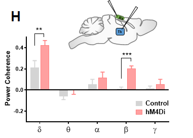
Multielectrode recordings suggest so!
➡️ All hyperconnected areas show robustly increased δ power & phase coherence
➡️ δ power is linearly related to mean rsfMRI connectivity in all areas
So δ band coherence is a plausible driver of the observed fMRI over-synchronization
13/n
➡️ All hyperconnected areas show robustly increased δ power & phase coherence
➡️ δ power is linearly related to mean rsfMRI connectivity in all areas
So δ band coherence is a plausible driver of the observed fMRI over-synchronization
13/n

Interestingly, our recordings also showed increased δ coupling between polymodal thalamus and posterior cingulate (retrosplenial in mouse)
➡️If delta is a prominent correlate of rsfMRI over-synchronization, these two regions should then exhibit rsfMRI overconnectivity too
14/n
➡️If delta is a prominent correlate of rsfMRI over-synchronization, these two regions should then exhibit rsfMRI overconnectivity too
14/n
This may be important mechanistically as polymodal thalamus generates and propagates δ activity to cortex
(see below)
If this were the case
➡️ polymodal/unimodal rsfMRI profiles should be segregable
➡️ polymodal thalamus would be "overconnected" with large cortical areas
15/n
(see below)
If this were the case
➡️ polymodal/unimodal rsfMRI profiles should be segregable
➡️ polymodal thalamus would be "overconnected" with large cortical areas
15/n

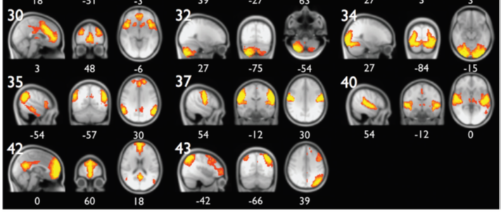
Using a clustering analysis we show this might be the case
➡️we found distinct poly/unimodal rsfMRI profiles
➡️polymodal (but not unimodal) thalamus exhibits rsfMRI overconnectivity with cortical areas
➡️ these include retrosplenial Ctx as predicted by ePHYS recordings
16/n
➡️we found distinct poly/unimodal rsfMRI profiles
➡️polymodal (but not unimodal) thalamus exhibits rsfMRI overconnectivity with cortical areas
➡️ these include retrosplenial Ctx as predicted by ePHYS recordings
16/n

This leads to a putative model whereby silencing of a cortical region leads to
➡️ reduced "direct" (e.g. ƴ) communication with its targets
➡️ & increased rsfMRI coupling mediated by co-varying slow oscillations (from polymodal thalamus and/or other remote rhythm generators)
17/n
➡️ reduced "direct" (e.g. ƴ) communication with its targets
➡️ & increased rsfMRI coupling mediated by co-varying slow oscillations (from polymodal thalamus and/or other remote rhythm generators)
17/n

These results may have important implications e.g.
for modelling ➡️rsfMRI connectivity is not always a monotonic indicator of interareal communication
for brain disorders ➡️overconnectivity in degenerative disorders may be indirect result of lower cortical output
18/n
for modelling ➡️rsfMRI connectivity is not always a monotonic indicator of interareal communication
for brain disorders ➡️overconnectivity in degenerative disorders may be indirect result of lower cortical output
18/n
This is it. Many thanks to twitterless
& freshly graduated 🍾 #Carola_Canella
#Federico_Rocchi, #Stefano_Panzeri, @danielgb_87 @GIurilli and all the collaborators at @IITalk
And to @BBRFoundation and @ERC_Research for generous funding. Comments/suggestions are welcome
19/n
& freshly graduated 🍾 #Carola_Canella
#Federico_Rocchi, #Stefano_Panzeri, @danielgb_87 @GIurilli and all the collaborators at @IITalk
And to @BBRFoundation and @ERC_Research for generous funding. Comments/suggestions are welcome
19/n