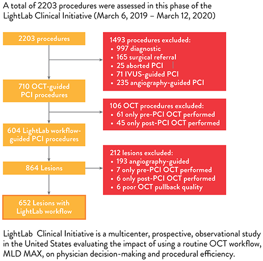
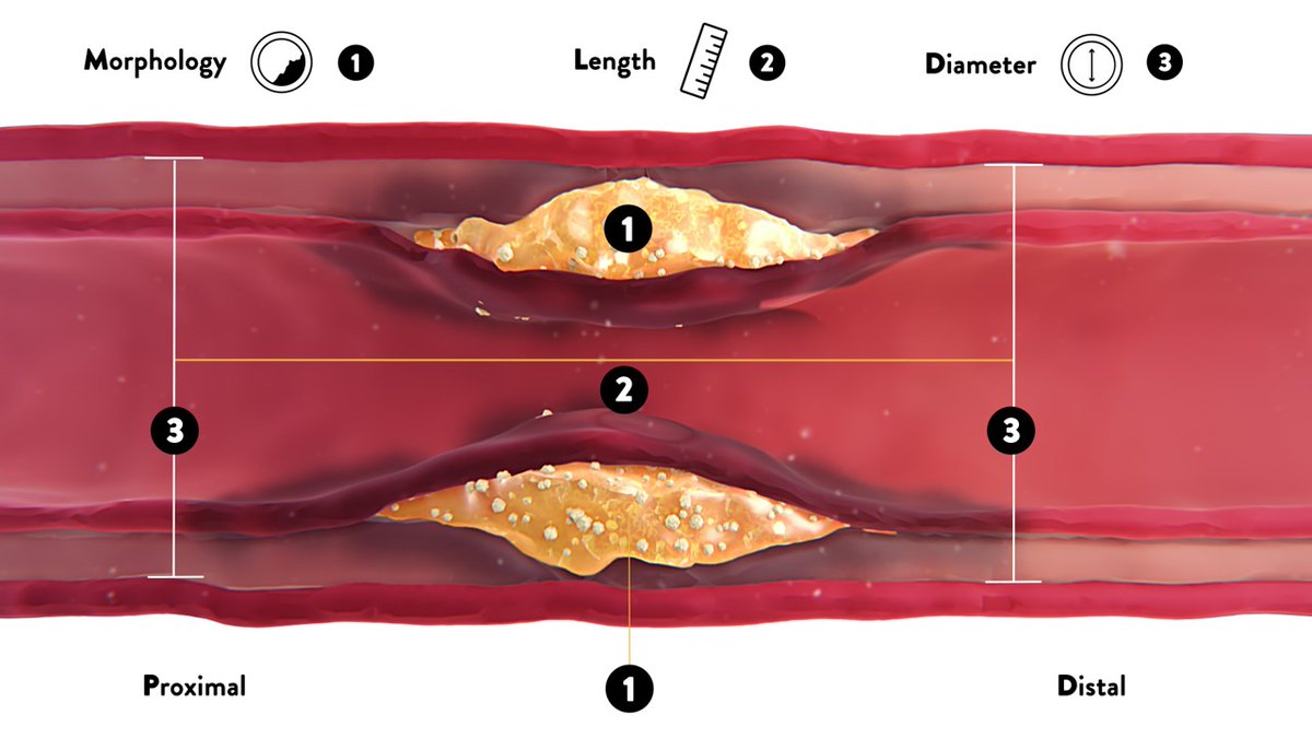
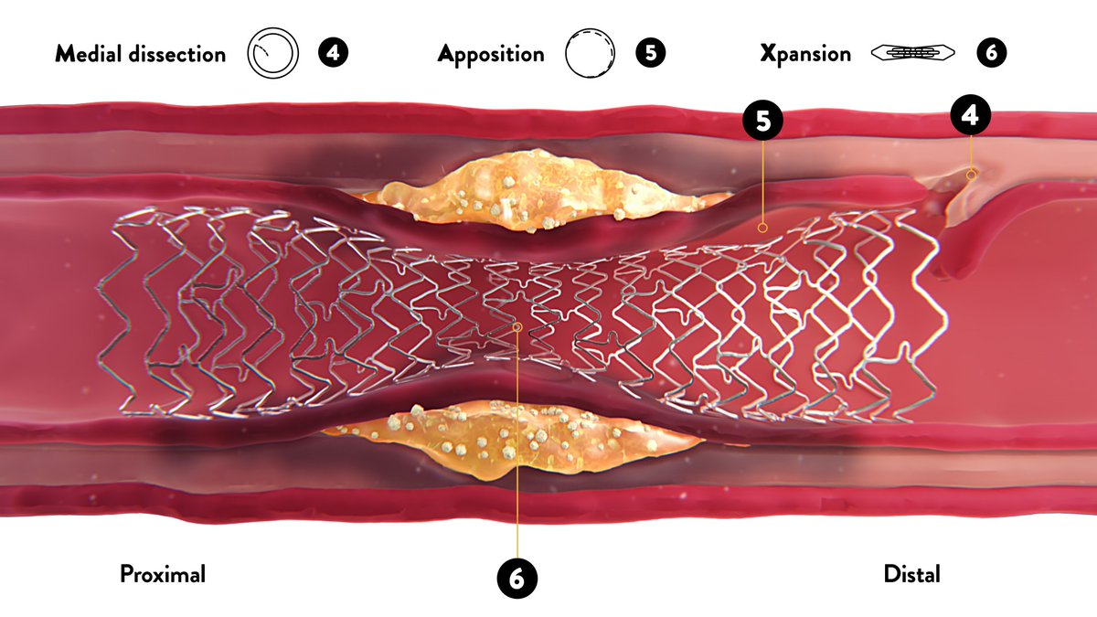
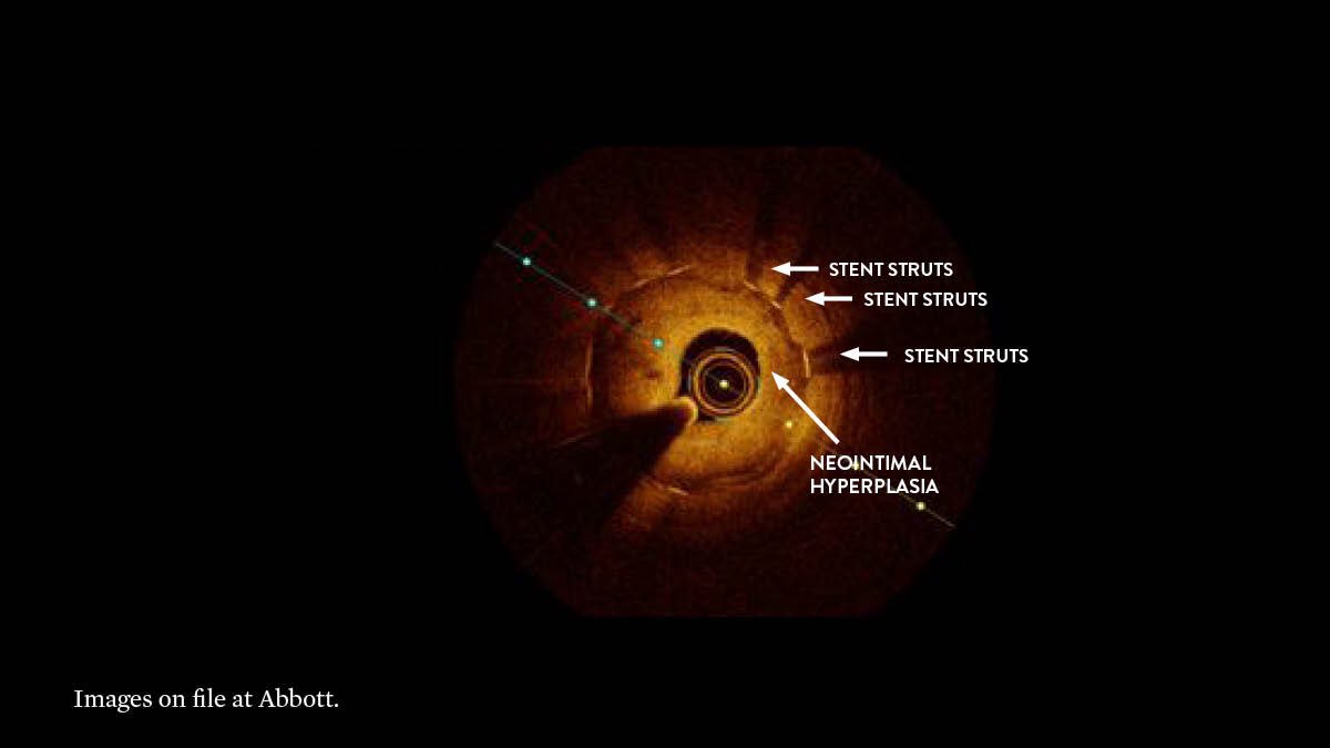
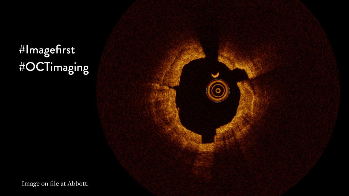
Today we discuss the 1st step in the OCT-guided PCI workflow #MLDMAX: Morphology (M)
At the end, assess your morphology skills w/ a quiz.
Important Safety Info: bit.ly/2VIph7r
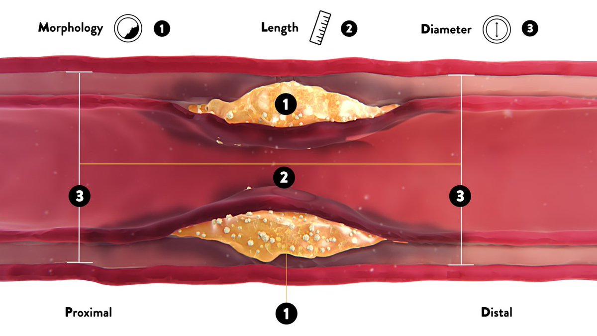
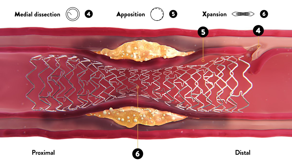
In the LightLab Clinical Initiative, #OCTimaging changed physician’s angiographic assessment of lesion morphology in 48% of lesions or about 1 out of every 2 lesions.
#imagefirst
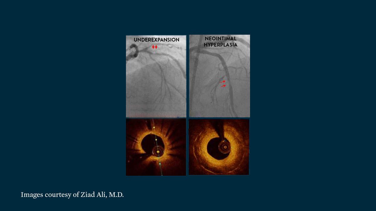
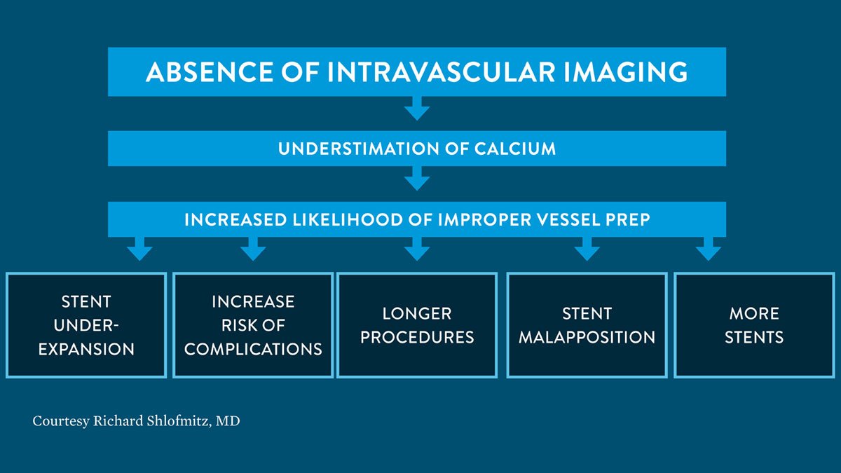
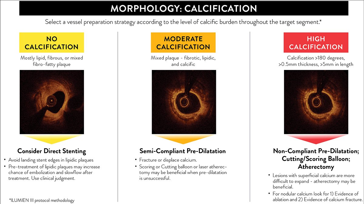
Calcified lesions are at an increased risk for stent underexpansion which is established as a predictor of stent failure, including stent thrombosis & restenosis.*
*Raber et al: bit.ly/RaberECD, SCAI: bit.ly/SCAI_oPCI
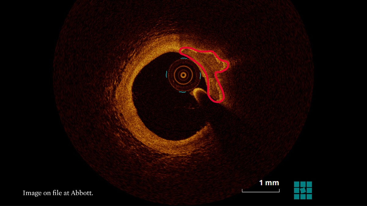
OCT can penetrate calcium and measure its thickness more accurately than IVUS.*
*Fujino et al bit.ly/FujinoCs, Raber et al bit.ly/RaberECS
#imagefirst #imagelast
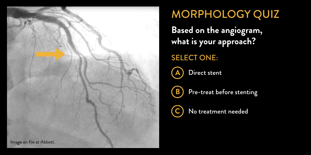
Check your skills in a poll below and RT to have your colleagues join!
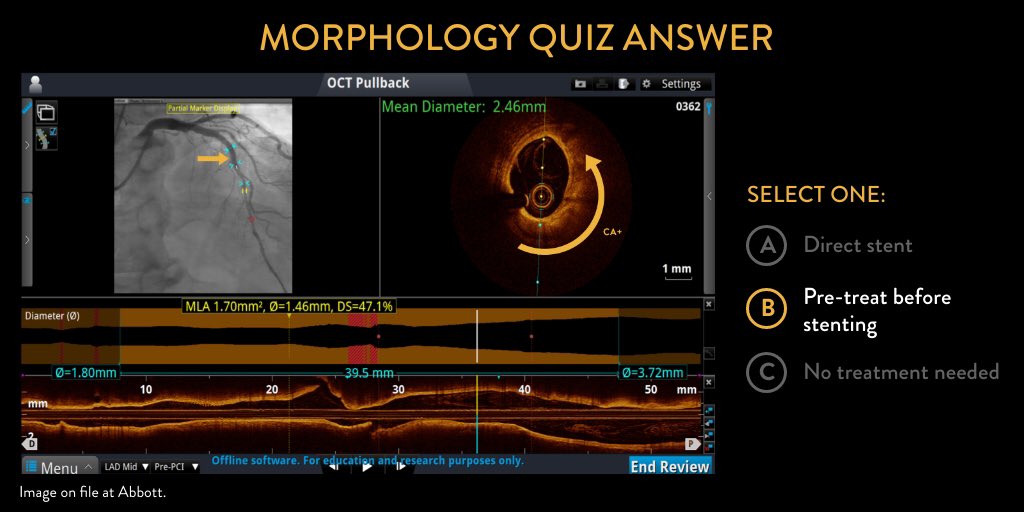
Based on this angiogram, what is your approach?
Important Safety Info: bit.ly/2VIph7r
OCT can assess lesion morphology to inform vessel prep strategy & develop a treatment approach.
Important Safety Info: bit.ly/2VIph7r
Experience the OCT difference, request an Abbott Sales Rep today: abbo.tt/3jRCJhh
Join us next Tuesday to discuss length and diameter in the #MLDMAX steps.
Important Safety Info: bit.ly/2VIph7r


