1/Welcome to our second OCTober #Tweetorial 🎃🤓. We’ll talk about diagnosing and treating calcific plaque with #OCTImaging. #imagefirst
Important Safety Info: abbo.tt/2GyBrZ1
Important Safety Info: abbo.tt/2GyBrZ1
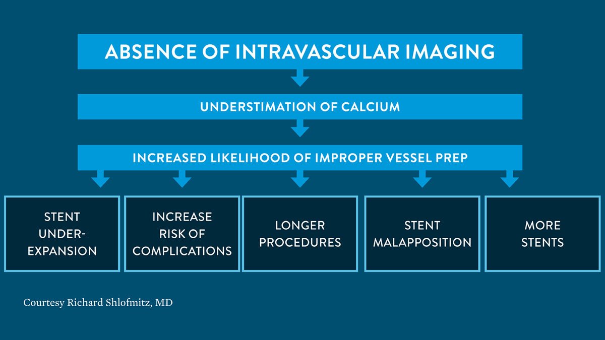
2/The absence of intravascular imaging may lead to underestimation of calcium and improper diagnosis, as seen in the chart below, courtesy of Richard Shlofmitz, MD. #OCTImaging #imagefirst
Important Safety Info: abbo.tt/2GyBrZ1
Important Safety Info: abbo.tt/2GyBrZ1
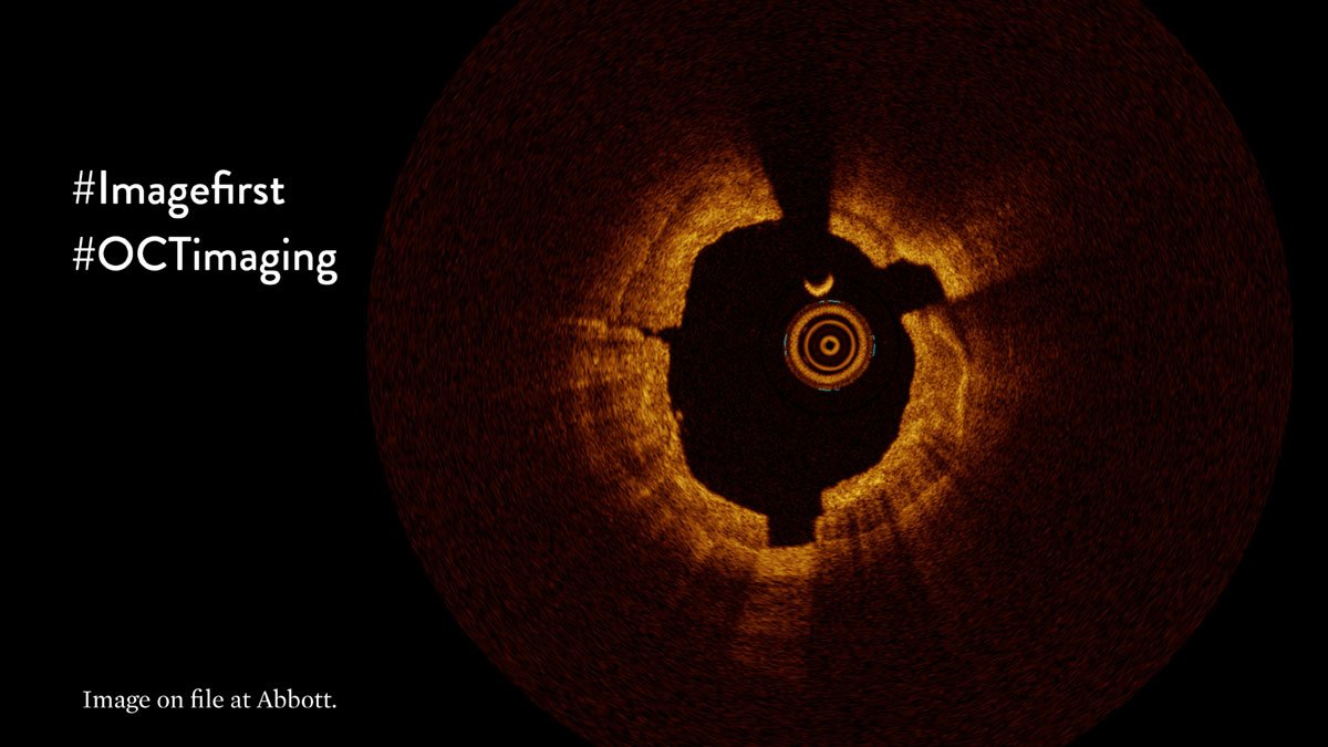
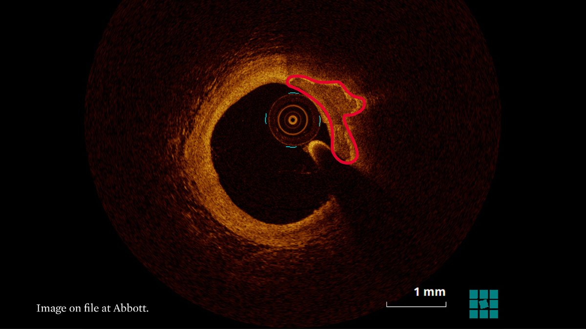
3/Calcific plaque is characterized as having low attenuation (light penetrates deep, can see tissue) & clear, delineated edges (appear as “islands” or “rocks”). In this image, calcific plaque is observed from 12 o’clock to 3 o’clock.
Important Safety Info: abbo.tt/2GyBrZ1
Important Safety Info: abbo.tt/2GyBrZ1
4/When diagnosing calcium, it’s important❗️ to differentiate between (A) deep, (B) superficial and (C) nodular calcium--as seen in these OCT images--to choose the right treatment strategy. #OCTImaging #imagefirst
Important Safety Info: abbo.tt/2GyBrZ1
Important Safety Info: abbo.tt/2GyBrZ1
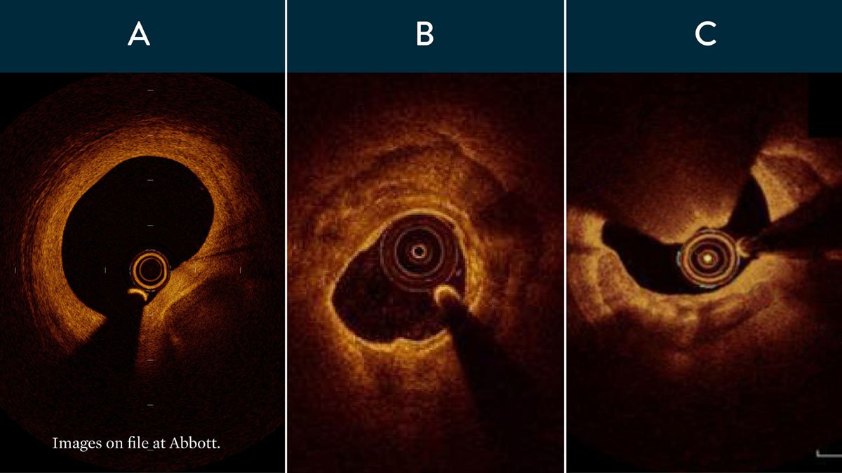
5/Let’s review how each calcium type appears on OCT.
Deep calcium appears furthest away from the lumen separated by a thick layer. #OCTImaging #imagefirst
Important Safety Info: abbo.tt/2GyBrZ1
Deep calcium appears furthest away from the lumen separated by a thick layer. #OCTImaging #imagefirst
Important Safety Info: abbo.tt/2GyBrZ1
6/Superficial calcium appears close to the lumen. #OCTImaging #imagefirst
Important Safety Info: abbo.tt/2GyBrZ1
Important Safety Info: abbo.tt/2GyBrZ1
7/Nodular calcium extends into the lumen. #OCTImaging #imagefirst
Important Safety Info: abbo.tt/2GyBrZ1
Important Safety Info: abbo.tt/2GyBrZ1
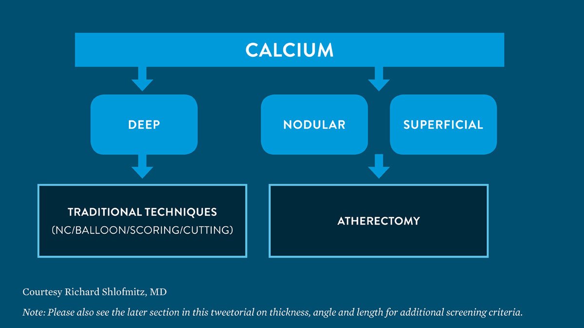
8/ By accessing the degree of calcium burden, you can identify the right pretreatment strategy. Use this handy chart developed by Richard Shlofmitz,
MD. #OCTImaging #imagefirst
Important Safety Info: abbo.tt/2GyBrZ1
MD. #OCTImaging #imagefirst
Important Safety Info: abbo.tt/2GyBrZ1
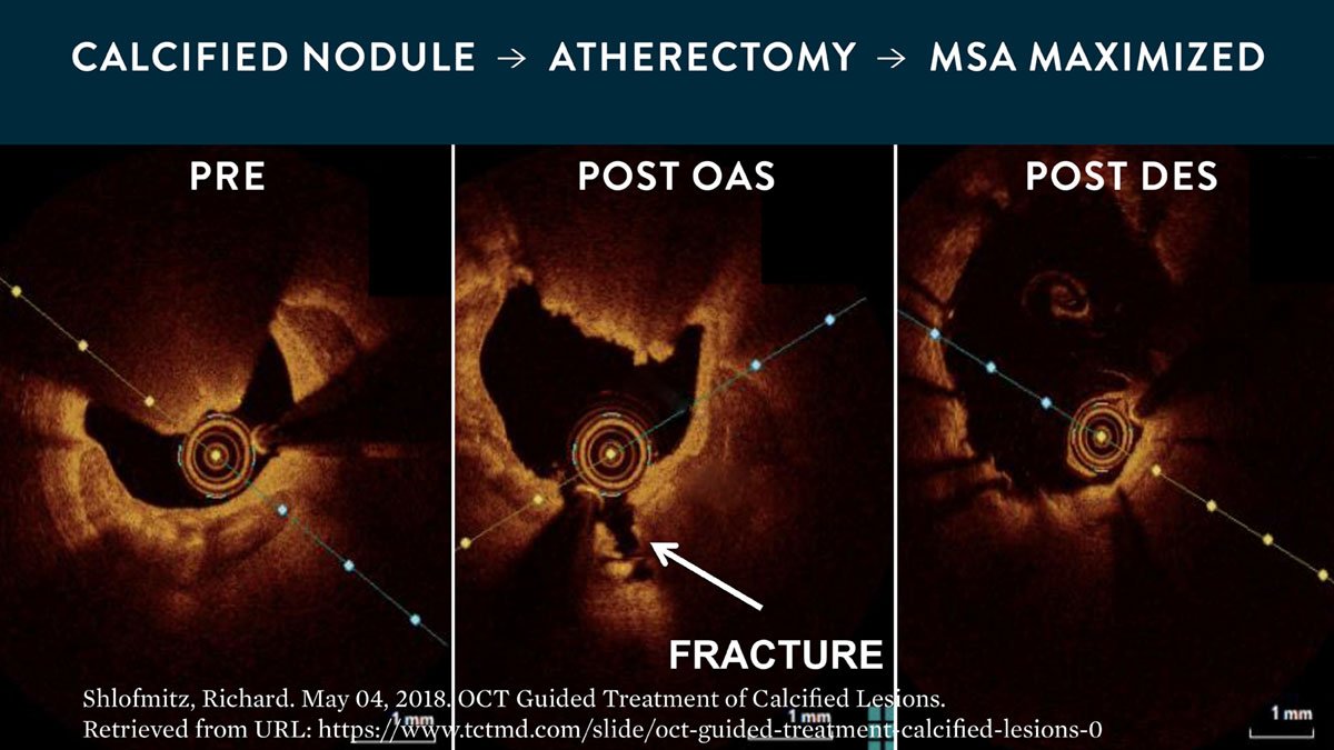
9/During atherectomy, OCT helps visualize fractures in and the reduction of calcium to guide further treatment decisions. #OCTImaging #imagefirst
Important Safety Info: abbo.tt/2GyBrZ1
Important Safety Info: abbo.tt/2GyBrZ1
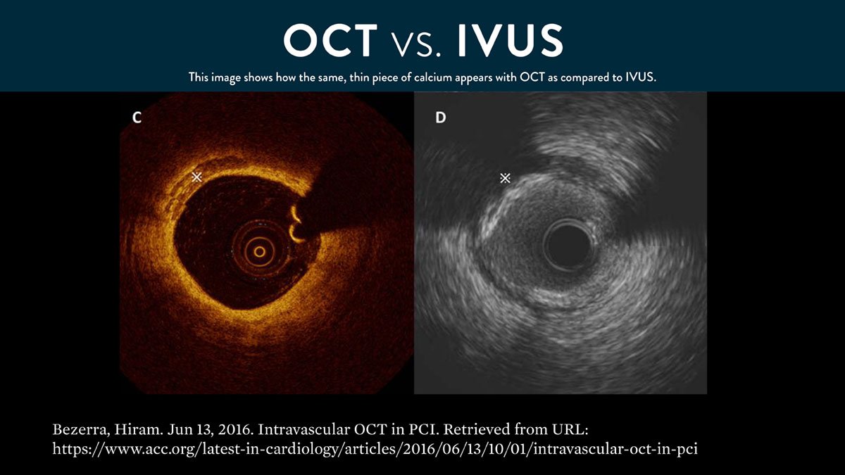
10/ #OCTimaging helps you realize the thickness of the calcium as compared to IVUS. OCT is light-based, it penetrates and surrounds calcium; IVUS is sound-based, sound waves bounce off calcium & create a dark shadow, seen below.
Important Safety Info: abbo.tt/2GyBrZ1
Important Safety Info: abbo.tt/2GyBrZ1
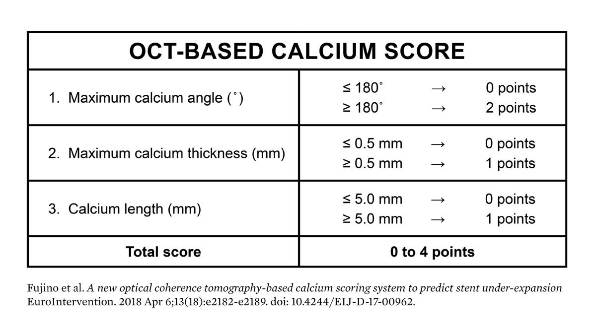
11/Measuring calcium depth w/ an OCT-based calcium score algorithm can help identify calcific lesions that would benefit from plaque modification before stent implantation, concluded in Fujino et al study: bit.ly/2pJ0LRh
Important Safety Info: abbo.tt/2GyBrZ1
Important Safety Info: abbo.tt/2GyBrZ1
12/This algorithm looks at calcium thickness, calcium angle and calcium length. Lesions at risk of stent under-expansion have a calcium score of 4, based on this algorithm.
➡️ Thickness >0.5 mm
➡️ Angle >180
➡️ Length >5 mm
Important Safety Info: abbo.tt/2GyBrZ1
➡️ Thickness >0.5 mm
➡️ Angle >180
➡️ Length >5 mm
Important Safety Info: abbo.tt/2GyBrZ1