Sanjiv Shah Profile picture
15 Dec, 12 tweets, 3 min read
#HFpEF pearl of the day: Bike stress echo is very helpful in the evaluation of HFpEF. In 1 test you can diagnose HFpEF (E/e') and evaluate for CAD (WMA), health of the LA (Δ LA strain), LV contractile reserve (Δ LV strain), dynamic MR, and dynamic LV outflow tract obstruction.
If RV free wall strain goes down with exercise, could be a sign of dynamic pulmonary vasoconstriction during exercise (i.e., ⬆️PVR with exercise) or intrinsic RV dysfunction.
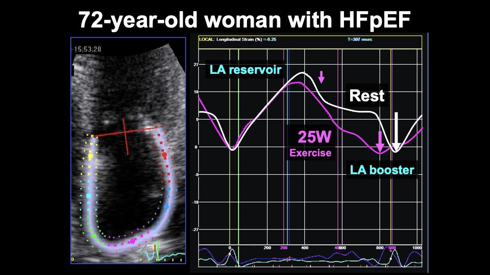
Here is an example. 72 yo woman w/HFpEF. With just 25W exercise, E velocity goes way up. And a' velocity goes down dramatically, indicative of LA contractile dysfunction and ⬆️LV end-diastolic stiffness. Image
Here is what happens to LA reservoir strain in the same patient. LA booster strain goes down at 25W of exercise. Conduit strain also goes down. LA reservoir strain = booster + conduit strains therefore also ⬇️. Image
Here is another patient with HFpEF who developed significant PAH superimposed (CpcPH) due to systemic sclerosis. Here is baseline echo:
TAPSE increased from 1.8 cm at rest to 2.5 cm at peak stress: Image
RV s' velocity increased from 13 cm/s to 19 cm/s at peak stress: Image
How did this patient's RV systolic function respond to exercise?
Here is the 2D echo (A4c view) at peak stress:
Answer: RV systolic function ⬇️ with exercise. TAPSE and RV s' velocity are unreliable because of tethering effect of the LV, which gets hyperdynamic during exercise, "pulling" the RV longitudinally in the absence of ⬆️RV contractility. Translational motion also problematic. Image
Unlike TAPSE and s' velocity, RV free wall strain worsens with exercise, indicating ⬇️RV systolic function. The RV dysfunction is clear on 2D echo w/exercise, so strain not necessarily needed, but helpful to show why it's better than displacement (TAPSE) and velocity (RV s').
RV free wall strain analysis can be helpful for more subtle cases of RV dysfunction is there is a question about whether RV is augmenting appropriately with exercise.

• • •

Missing some Tweet in this thread? You can try to force a refresh
 

Keep Current with Sanjiv Shah

Sanjiv Shah Profile picture

Stay in touch and get notified when new unrolls are available from this author!

Read all threads

This Thread may be Removed Anytime!

PDF

Twitter may remove this content at anytime! Save it as PDF for later use!

Try unrolling a thread yourself!

how to unroll video
  1. Follow @ThreadReaderApp to mention us!

  2. From a Twitter thread mention us with a keyword "unroll"
@threadreaderapp unroll

Practice here first or read more on our help page!

More from @HFpEF

18 Dec
#HFpEF pearl of the day: Always look at echo LVOT VTI when eval. HFpEF pts. Normal 18-22 cm at HR 60-100. ⬆️LVOT VTI: look for NASH, anemia, cirrhosis, AVMs, etc. If ⬇️LVOT VTI: infiltrative/restrictive CM, constriction, LA failure, PAH, RV failure, obstructive lung dz, MS, etc.
If ⬆️LVOT, measure volumes/dimensions of all 4 cardiac chambers to look for enlargement. Case: 31 yo woman previously healthy presents with dyspnea, leg swelling, BNP 166 pg/ml. Training for a marathon. Urine pregnancy test negative.
Echo shows normal LVEF (65%). E/e’ = 6. Lateral e’ velocity = 15 cm/s. LV dilated (LVEDVI = 85 ml/m2) and LA dilated (LA volume index = 50 ml/m2). ⬆️LVOT VTI = 30 cm. Image
Read 6 tweets
16 Dec
#HFpEF pearl of the day: In HFpEF pts with #CardioMEMS devices, bike stress echo + continuous CardioMEMS PA pressure recording can help evaluate underlying pathophysiologic abnormalities.
Case: 71 yo woman w/apical HCM, AF s/p ablation, pacemaker, HTN, CKD with cardiorenal syndrome and PH-HFpEF presents with worsening overload. Echo shows preserved LVEF with apical HCM, PASP 85 mmHg, RAP 15 mmHg, RVOT PW notching consistent with ⬆️PVR.
Image
Read 17 tweets
12 Dec
Answer to today's case: the pt had a very stiff LA, poss. due to AF ablation. But stiff LA alone cannot cause big V waves. Sympathetic activation➡️splanchnic vasoconstriction w/redistribution of volume from gut/liver➡️lungs/heart with minimal exertion + stiff LA = big V waves.
This pt also had significant coronary microvascular dysfxn. Exercise➡️myocardial ischemia➡️LV diastolic dysfunction➡️increased load on LA. I tried everything to treat him, nothing worked for 7 years. And then he got an IASD as part of the @corviamedical REDUCE LAP-HF I trial.
Huge improvement in symptoms, now NYHA class I-II.
Read 4 tweets
11 Dec
Case: 64 yo man w/HFpEF, HTN, obesity, CAD, AF s/p ablation, NYHA 3. Invasive hemodynamics shown below. PCWP 11 at rest, ⬆️ to 32 with V waves up to 75 with 20W bike exercise. Only 1+ MR at rest/exercise. What caused the severe elevation in PCWP and V waves with minimal exercise?
Read 5 tweets
11 Dec
#HFpEF pearl of the day: There are both "slow" and "fast" mechanisms of congestion in HFpEF patients. The "fast" mechanism is mediated by splanchnic vasoconstriction, which also may have implications for cardiorenal syndrome.
Read 4 tweets
9 Dec
Case: 74-year-old woman with HTN, obesity (BMI 46 kg/m2) with dyspnea, leg edema, ⬆️JVP, BNP = 28 pg/ml, LVEF 60%. What is the cause of HFpEF and abnormal septal motion?
Invasive hemodynamics: RA 18, PA 44/20 (mean 28), PCWP 20, CO 5.1 L/min. Image
Answer: Obesity HFpEF phenotype with severe pericardial, epicardial fat over the RV causing pericardial constraint (not constrictive pericarditis--pericardium was normal). See: ahajournals.org/doi/10.1161/CI… Image
Read 9 tweets

Did Thread Reader help you today?

Support us! We are indie developers!


This site is made by just two indie developers on a laptop doing marketing, support and development! Read more about the story.

Become a Premium Member ($3/month or $30/year) and get exclusive features!

Become Premium

Too expensive? Make a small donation by buying us coffee ($5) or help with server cost ($10)

Donate via Paypal Become our Patreon

Thank you for your support!

Follow Us on Twitter!