Sanjiv Shah Profile picture
16 Dec, 17 tweets, 5 min read
#HFpEF pearl of the day: In HFpEF pts with #CardioMEMS devices, bike stress echo + continuous CardioMEMS PA pressure recording can help evaluate underlying pathophysiologic abnormalities.
Case: 71 yo woman w/apical HCM, AF s/p ablation, pacemaker, HTN, CKD with cardiorenal syndrome and PH-HFpEF presents with worsening overload. Echo shows preserved LVEF with apical HCM, PASP 85 mmHg, RAP 15 mmHg, RVOT PW notching consistent with ⬆️PVR.
Invasive hemodynamics showed RA pressure 25 mmHg, PA pressure 92/31 (mean 54), PCWP 31 mmHg with V waves up to 60 mmHg.
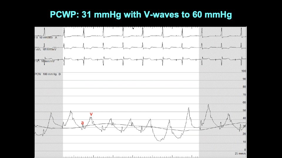
CardioMEMS placed showing good agreement with invasive hemodynamics:
She was admitted and underwent IV diuresis. Still very dyspneic. Exercise echo with continuous CardioMEMS pressure recordings done while inpatient. She could only exercise on bike to 15W. ⬆️PA pulse pressure, minimal HR increase.
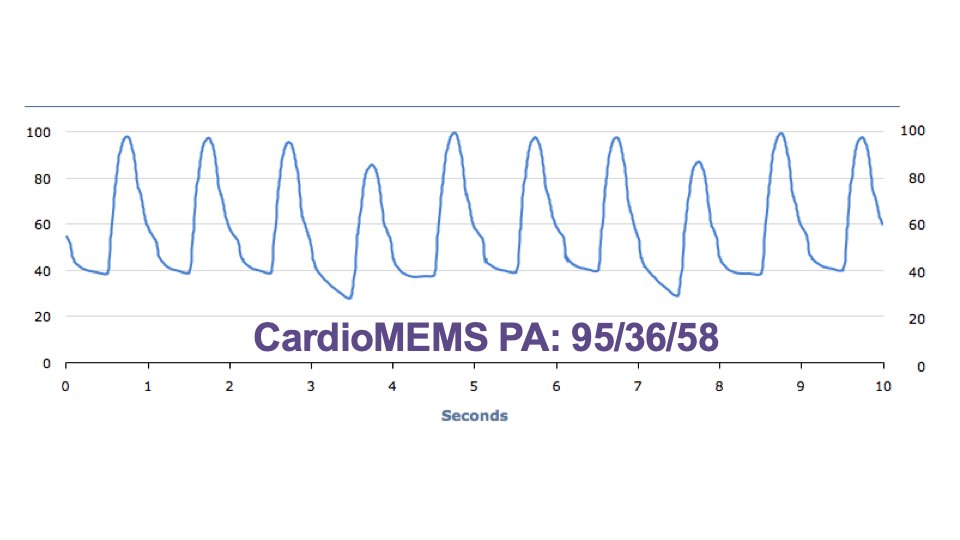
Here are the CardioMEMS tracings captured during the bike stress echo:
Mitral inflow E velocity went down with exercise, suggesting underfilling of the LA:
LVOT VTI did not augment with exercise suggesting fixed stroke volume.
She had a wrist watch arterial tonometry device as well:
Systemic pulse pressure went down with exercise:
At 15W exercise, PA pulse pressure went up, and RV free wall strain got worse.
Meanwhile, at 15W exercise, LV longitudinal strain got better (augmented).
Based on the bike stress echo + CardioMEMS it seemed that SV fixed and inadequate HR, so CO was impaired. Blood trapped in the pulmonary circulation w/⬆️ PA pulse pressure and underfilling of LA/LV. Rx'd by ⬆️ basal HR on pacemaker, ⬆️ rate responsiveness: improved symptoms.
Ideally we could’ve done something to ⬆️pulmonary blood flow during exercise so that she could better ⬆️SV but unfortunately no options available. We continued to diurese and Rx her w/hemodynamic-guided mgmt but unfortunately she died ~1 yr later due to progressive renal failure.

• • •

Missing some Tweet in this thread? You can try to force a refresh
 

Keep Current with Sanjiv Shah

Sanjiv Shah Profile picture

Stay in touch and get notified when new unrolls are available from this author!

Read all threads

This Thread may be Removed Anytime!

PDF

Twitter may remove this content at anytime! Save it as PDF for later use!

Try unrolling a thread yourself!

how to unroll video
  1. Follow @ThreadReaderApp to mention us!

  2. From a Twitter thread mention us with a keyword "unroll"
@threadreaderapp unroll

Practice here first or read more on our help page!

More from @HFpEF

18 Dec
#HFpEF pearl of the day: Always look at echo LVOT VTI when eval. HFpEF pts. Normal 18-22 cm at HR 60-100. ⬆️LVOT VTI: look for NASH, anemia, cirrhosis, AVMs, etc. If ⬇️LVOT VTI: infiltrative/restrictive CM, constriction, LA failure, PAH, RV failure, obstructive lung dz, MS, etc.
If ⬆️LVOT, measure volumes/dimensions of all 4 cardiac chambers to look for enlargement. Case: 31 yo woman previously healthy presents with dyspnea, leg swelling, BNP 166 pg/ml. Training for a marathon. Urine pregnancy test negative.
Echo shows normal LVEF (65%). E/e’ = 6. Lateral e’ velocity = 15 cm/s. LV dilated (LVEDVI = 85 ml/m2) and LA dilated (LA volume index = 50 ml/m2). ⬆️LVOT VTI = 30 cm. Image
Read 6 tweets
15 Dec
#HFpEF pearl of the day: Bike stress echo is very helpful in the evaluation of HFpEF. In 1 test you can diagnose HFpEF (E/e') and evaluate for CAD (WMA), health of the LA (Δ LA strain), LV contractile reserve (Δ LV strain), dynamic MR, and dynamic LV outflow tract obstruction.
If RV free wall strain goes down with exercise, could be a sign of dynamic pulmonary vasoconstriction during exercise (i.e., ⬆️PVR with exercise) or intrinsic RV dysfunction.
Here is an example. 72 yo woman w/HFpEF. With just 25W exercise, E velocity goes way up. And a' velocity goes down dramatically, indicative of LA contractile dysfunction and ⬆️LV end-diastolic stiffness. Image
Read 12 tweets
12 Dec
Answer to today's case: the pt had a very stiff LA, poss. due to AF ablation. But stiff LA alone cannot cause big V waves. Sympathetic activation➡️splanchnic vasoconstriction w/redistribution of volume from gut/liver➡️lungs/heart with minimal exertion + stiff LA = big V waves.
This pt also had significant coronary microvascular dysfxn. Exercise➡️myocardial ischemia➡️LV diastolic dysfunction➡️increased load on LA. I tried everything to treat him, nothing worked for 7 years. And then he got an IASD as part of the @corviamedical REDUCE LAP-HF I trial.
Huge improvement in symptoms, now NYHA class I-II.
Read 4 tweets
11 Dec
Case: 64 yo man w/HFpEF, HTN, obesity, CAD, AF s/p ablation, NYHA 3. Invasive hemodynamics shown below. PCWP 11 at rest, ⬆️ to 32 with V waves up to 75 with 20W bike exercise. Only 1+ MR at rest/exercise. What caused the severe elevation in PCWP and V waves with minimal exercise?
Read 5 tweets
11 Dec
#HFpEF pearl of the day: There are both "slow" and "fast" mechanisms of congestion in HFpEF patients. The "fast" mechanism is mediated by splanchnic vasoconstriction, which also may have implications for cardiorenal syndrome.
Read 4 tweets
9 Dec
Case: 74-year-old woman with HTN, obesity (BMI 46 kg/m2) with dyspnea, leg edema, ⬆️JVP, BNP = 28 pg/ml, LVEF 60%. What is the cause of HFpEF and abnormal septal motion?
Invasive hemodynamics: RA 18, PA 44/20 (mean 28), PCWP 20, CO 5.1 L/min. Image
Answer: Obesity HFpEF phenotype with severe pericardial, epicardial fat over the RV causing pericardial constraint (not constrictive pericarditis--pericardium was normal). See: ahajournals.org/doi/10.1161/CI… Image
Read 9 tweets

Did Thread Reader help you today?

Support us! We are indie developers!


This site is made by just two indie developers on a laptop doing marketing, support and development! Read more about the story.

Become a Premium Member ($3/month or $30/year) and get exclusive features!

Become Premium

Too expensive? Make a small donation by buying us coffee ($5) or help with server cost ($10)

Donate via Paypal Become our Patreon

Thank you for your support!

Follow Us on Twitter!