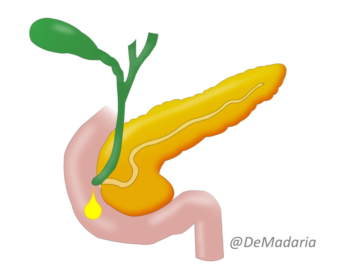
Cross-sectional imaging often reveals unexpected pancreatic cystic lesions, it is a frequent clinical problem, Should we observe or remove it? What's the diagnosis? Is our patient in danger of malignancy?
Don’t miss this @aegastro @my_ueg #EducAEG #UEGambassador twitter thread
Don’t miss this @aegastro @my_ueg #EducAEG #UEGambassador twitter thread

Importance of Pancreatic Cystic Neoplasms (PCN):
Most are asymptomatic at diagnosis, frequency increases with age
Symptoms: acute pancreatitis (Wirsung obstructed by the cyst or mucus), pain, obstructive chronic pancreatitis, jaundice
> symptoms, >malignancy risk!
Most are asymptomatic at diagnosis, frequency increases with age
Symptoms: acute pancreatitis (Wirsung obstructed by the cyst or mucus), pain, obstructive chronic pancreatitis, jaundice
> symptoms, >malignancy risk!

Classification of PCN:
Mucinous: intraductal papillary mucinous neop. and mucinous cystic neop.
Nonmucinous: serous cystic neoplasm, solid pseudopapillary neoplasm and cystic neuroendocrine tumours
Endoderm- derived columnar epithelium is characteristic for mucinous lesions
👇
Mucinous: intraductal papillary mucinous neop. and mucinous cystic neop.
Nonmucinous: serous cystic neoplasm, solid pseudopapillary neoplasm and cystic neuroendocrine tumours
Endoderm- derived columnar epithelium is characteristic for mucinous lesions
👇

Intraductal papillary mucinous neoplasms (IPMN)
Characterized by papillary proliferation+mucus production. It may involve Wirsung (becomes dilated) and/or branch ducts (cysts connected to the ductal system). It may evolve to pancreatic cancer particularly if Wirsung is involved

Characterized by papillary proliferation+mucus production. It may involve Wirsung (becomes dilated) and/or branch ducts (cysts connected to the ductal system). It may evolve to pancreatic cancer particularly if Wirsung is involved


IPMN subtypes :
Intestinal: main duct, head, 40%->coloid/tubular adenoca
Pancreatobiliary: main duct,head, 68%->tubular adenoca
Oncocytic: rare, nodules,50%-> coloid/tubular adenoca
Gastric: most frequent, branch-type, uncinate, 10%->tubular adenoca
nature.com/articles/s4157…
Intestinal: main duct, head, 40%->coloid/tubular adenoca
Pancreatobiliary: main duct,head, 68%->tubular adenoca
Oncocytic: rare, nodules,50%-> coloid/tubular adenoca
Gastric: most frequent, branch-type, uncinate, 10%->tubular adenoca
nature.com/articles/s4157…
IPMN: risk factors for malignancy
Main duct involvement (60% in resected specimens vs 10 to 30% in resected side branch IPMNs), specially>1cm
Contrast-enhanced mural nodules
Size>3-4cm
Symptoms
Pts at risk of PDAC even in other regions of the gland without involvement
👇👇👇
Main duct involvement (60% in resected specimens vs 10 to 30% in resected side branch IPMNs), specially>1cm
Contrast-enhanced mural nodules
Size>3-4cm
Symptoms
Pts at risk of PDAC even in other regions of the gland without involvement
👇👇👇

Intraductal papillary mucinous neoplasms -> management: follow these guidelines:
European guidelines 2018 @Gut_BMJ @chiaro_del @MarcBesselink doi.org/10.1136/gutjnl…
Fukuoka 2017 @pancreatology@SalviaRobi sciencedirect.com/science/articl…
European guidelines 2018 @Gut_BMJ @chiaro_del @MarcBesselink doi.org/10.1136/gutjnl…
Fukuoka 2017 @pancreatology@SalviaRobi sciencedirect.com/science/articl…
Mucinous Cystic Neoplasms (MCN)
Characterized by mucinous epithelium and ovarian-type stroma, in body/tail
It is described as macrocystic, septated cyst with small number of cavities, it may have eccentric calcifications, no connection to ductal system
95% women, 5-7th decades
Characterized by mucinous epithelium and ovarian-type stroma, in body/tail
It is described as macrocystic, septated cyst with small number of cavities, it may have eccentric calcifications, no connection to ductal system
95% women, 5-7th decades

Management of MCN according to the European guidelines: a conservative approach is recommended for asymptomatic MCN measuring <40 mm without an enhancing nodule
doi.org/10.1136/gutjnl…
@chiaro_del @MarcBesselink @Gut_BMJ
doi.org/10.1136/gutjnl…
@chiaro_del @MarcBesselink @Gut_BMJ
Serous cystic neoplasm (SCN). Cuboidal epithelium without dysplasia
70% women, 5-7th decades, NON-MUCINOUS solitary lesion
Classic SCN is microcystic (multiple small cysts, honeycomb-like) but can be macrocystic or solid. A central scar or calcification can be present


70% women, 5-7th decades, NON-MUCINOUS solitary lesion
Classic SCN is microcystic (multiple small cysts, honeycomb-like) but can be macrocystic or solid. A central scar or calcification can be present


SCN management: remove only if symptoms, for example this case from @Dhgua, the patient had jaundice due to a a massive SCN, a Whipple procedure was performed 

Cystic neuroendocrine tumor
It is a pancreatic NET with a central cystic changes. Solitary lesion, 5-6th decades, frequently with wall contrast enhancement, 10% malignant potential

It is a pancreatic NET with a central cystic changes. Solitary lesion, 5-6th decades, frequently with wall contrast enhancement, 10% malignant potential


Cystic neuroendocrine tumor management: asymptomatic and <2 cm you may follow the patient doi.org/10.1159/000443…
It seems that these cystic NET are less aggressive than solid NET
It seems that these cystic NET are less aggressive than solid NET
Finally,solid pseudopapillary neoplasm
They have malignant potential(15%), >risk if >5 cm
Young women=90% (2-3rd decades),body/tail.Solid and cystic solitary masses, calcifications,often with intracystic bleeding.They can spread to the peritoneum or distant organs like the liver

They have malignant potential(15%), >risk if >5 cm
Young women=90% (2-3rd decades),body/tail.Solid and cystic solitary masses, calcifications,often with intracystic bleeding.They can spread to the peritoneum or distant organs like the liver


This twitter thread was based on:
doi.org/10.1038/s41575…
doi.org/10.1136/gutjnl…
doi.org/10.1016/j.suc.…
And Pancreatic cystic neoplasms, several articles from @UpToDate Editors: JR Saltzman S Grover Authors: Asif Khalid, MDKevin McGrath, MD uptodate.com
doi.org/10.1038/s41575…
doi.org/10.1136/gutjnl…
doi.org/10.1016/j.suc.…
And Pancreatic cystic neoplasms, several articles from @UpToDate Editors: JR Saltzman S Grover Authors: Asif Khalid, MDKevin McGrath, MD uptodate.com
If you liked this twitter thread, please retweet the first tweet and follow me! #PancreasTwitter
I hope you enjoyed it, it took me a lot of effort to do this! 😅
I hope you enjoyed it, it took me a lot of effort to do this! 😅
@drdalbir @BilalMohammadMD @KralJan @drkeithsiau @MZorniak @DCharabaty @RashidLui @SunilAminMD @SanchezLunaMD @stevenbollipo @Samir_Grover @RishadJkhan @drmoutaz @RodriguezParra_
• • •
Missing some Tweet in this thread? You can try to
force a refresh


