On today's episode of “PUTTING YOUR IGNORANCE ON DISPLAY!” What does a partial- and full-thickness tear look like? Today, I had a sudden understanding/facepalming moment... @Retlouping@ShoulderGeek1@DrJN_SportsMed@JeremyLewisPT
Here was my thought, how can a full-thickness tear be small-medium-large when its FULL thickness? Full is full, how can it be small? This might be obvious to u...
Here was my internal image of what a partial and full-thickness tear looked like.
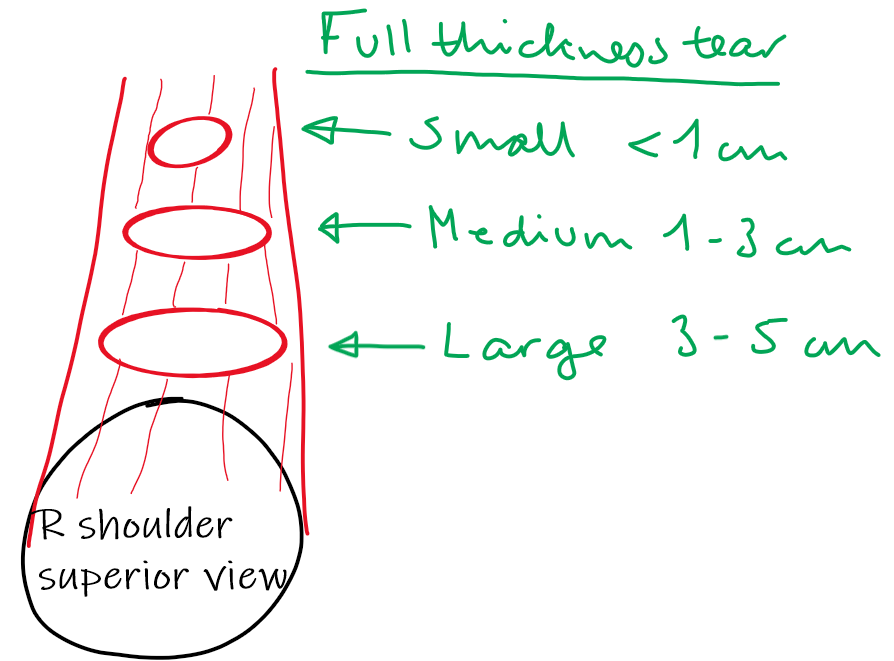
Of course, a full-thickness tear can look like the picture above, but here is an image of the “correct” way to look at it. The first picture is a superior view of the right shoulder with the supraspinatus. The second is an anterior view. The two images are of the same two tears.
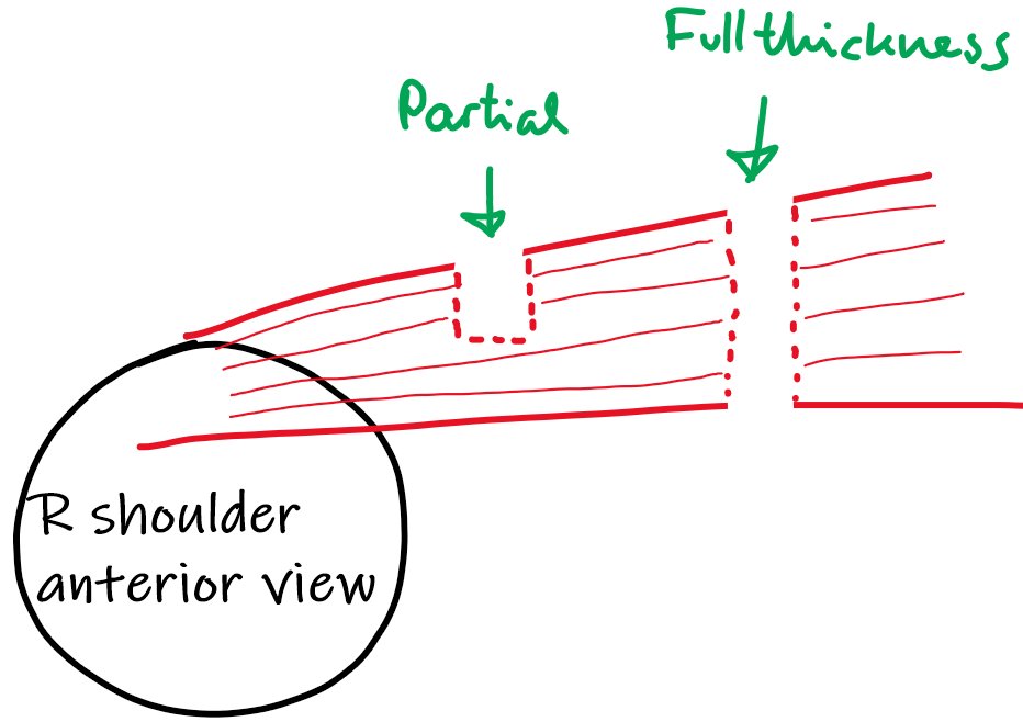
Full-thickness describes a HOLE through the FULL THICKNESS of the muscle belly. It describes a superior-to-inferior tear pattern, NOT an anterior-to-posterior tear pattern (like picture 1)
Now it becomes obvious how a full-thickness tear can be different sizes. It's just the diameter of the hole.
I would be very interested to hear if anyone had/have the same misunderstanding.
Also, praise for my painting skills would not go amiss 😁
My chiro friend is going over "IMPINGEMENT SYNDROME" of the shoulder at school. So I pointed him to these 4 articles. Might only be a colloquial term for some, but still... Let's put "impingement" behind us. We can and should do better!
1: The aim of the Ober test and the Modified Ober test is to assess the ITB.
2: The Ober test and Modified Ober test claims to assess ITB tightness
Let me present the coffin ⚰️
In the paper by Willett et al., 2016, they tested whether the ITB limits hip adduction during the Ober test and Modified Ober test. One would expect an INCREASE in hip adduction if the ITB was to be cut. pubmed.ncbi.nlm.nih.gov/26755689/
Catchy heading? Yes, it is a bit hyperbole. Now, on to the article.
As seen in the graph, the NHE out-performs the SLD, not only in overall muscle activation (y-axis), but also in every single muscle region (x-axis).
"absolute activity was substantially lower in both
muscles (ST = 72% vs 37%, BFlh = 64% vs 36%, of MVIC
on average in NHE vs SDL)."
Some muscles, we test at their "optimal" angle, whilst others we place at a less favourable angle for producing torque.
In the paper "Kinesiology of the hip - A focus on muscular action", by Neumann, Neumann has this to say about the abductor muscles: " Peak hip abduction torque occurs when the abductor muscles are nearly maximally elongated, in a position of 10 degrees of ❗️adduction❗️.
Excuse my (hopefully) catching and maybe slight provocative heading. I am a physiotherapy student who wants to embark on this exploratory journey with the hopeful help of more experienced minds.
In this thread, I want to propose that the anatomy of failure is the end-plate (EP), rather than the annulus fibrosus (AF). Hopefully, this might add some clinical value and a proposed explanation as to why some patients do worse in the long-term following a disc herniation.