OK #VExUS #POCUS enthusiasts, time for another case discussion.
Somebody asked if I ever recommend IV fluid in a patient with #VExUS 3.
Here is one example where I did.
1/ First, let's see the #physicalexam (#IMPOCUS) findings, then will tell about the case. #MedEd #Nephrology
Somebody asked if I ever recommend IV fluid in a patient with #VExUS 3.
Here is one example where I did.
1/ First, let's see the #physicalexam (#IMPOCUS) findings, then will tell about the case. #MedEd #Nephrology

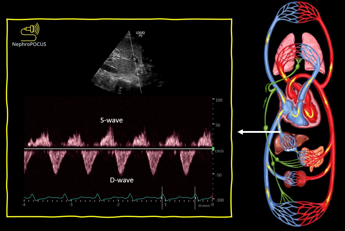
2/ So, hepatic shows D-only pattern👆
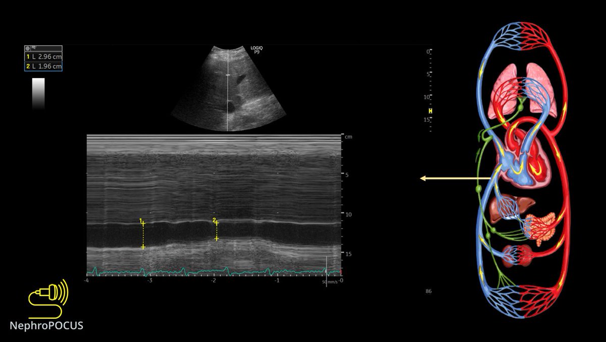
If we are doing #VExUS, IVC must be big. Here is the M-mode #POCUS 👇
If we are doing #VExUS, IVC must be big. Here is the M-mode #POCUS 👇
4/ D-only is severe congestion but its 👆better than the recent D-only we saw in a hyponatremic patient👇 Remember? (better in terms of how much cardiac cycle has venous flow) #POCUS 

5/ So, by definition, its already #VExUS grade 3 because 2 of the 3 veins we evaluate demonstrate severe flow changes. 

7/ This patient has h/o pulmonary hypertension. Remember, we discussed that hepatic and renal veins might never be normal in such cases but portal can normalize & helps when there is superimposed fluid overload?!
8/ Now, a few pictures of the pump.
RVOT Doppler #POCUS
V-shape is a little abnormal but no prominent notching appreciated unlike a recent case where I showed the 'W-pattern'
RVOT Doppler #POCUS
V-shape is a little abnormal but no prominent notching appreciated unlike a recent case where I showed the 'W-pattern'
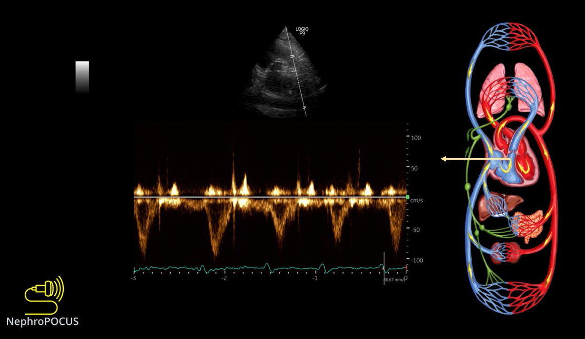
9/ For more information on RVOT Doppler #POCUS, you need to watch this video by hemodynamic master @khaycock2
vimeo.com/493079632
vimeo.com/493079632
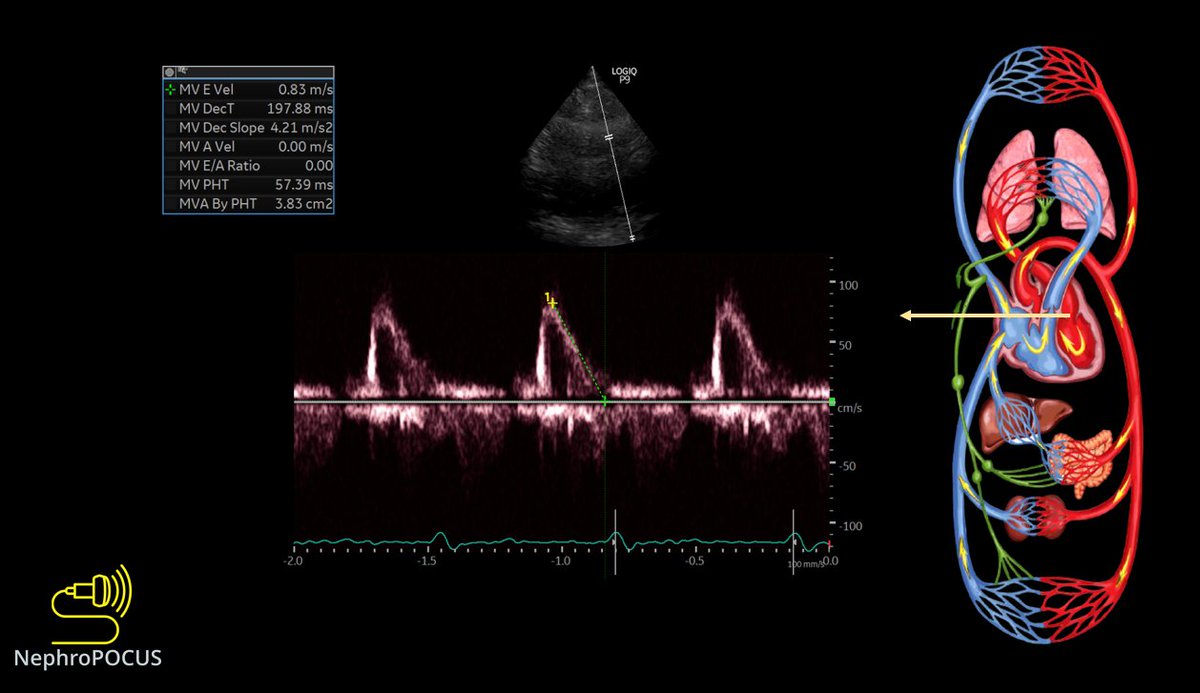
10/ Quick look at the left heart filling pressures using mitral inflow Doppler #POCUS and lateral annulus tissue Doppler.
E-wave deceleration time seems normal (an indicator of PCWP)
E-wave deceleration time seems normal (an indicator of PCWP)
12/ Lung #POCUS predominantly A-lines with B-lines in basal zones associated with irregular pleura. Likely chronic changes.
So why fluids and what's the clinical context?
So why fluids and what's the clinical context?
13/ Patient had AKI, likely secondary to ATN. While the serum creatinine is improving, developed post-ATN diuresis with an UO of ~5L in 24-hours.
Also had worsening hypernatremia with a calculated free water deficit of ~2 L + metabolic alkalosis.
Also had worsening hypernatremia with a calculated free water deficit of ~2 L + metabolic alkalosis.

14/ My recommendation was to give Half NS to replace at least half of the UO over the next 24 hours. Chloride containing fluid also helps with alkalosis.
That's it! I don't prefer free water 'flushes' through NG tube when significant amount of water has to be replaced.
That's it! I don't prefer free water 'flushes' through NG tube when significant amount of water has to be replaced.
15/ #VExUS crew for input/comments.
@ArgaizR @khaycock2 @ThinkingCC @RJonesSonoEM @katiewiskar @Scottiedoc1 @iceman_ex @curromir @KalagaraHari
@ArgaizR @khaycock2 @ThinkingCC @RJonesSonoEM @katiewiskar @Scottiedoc1 @iceman_ex @curromir @KalagaraHari
• • •
Missing some Tweet in this thread? You can try to
force a refresh


