Right side:
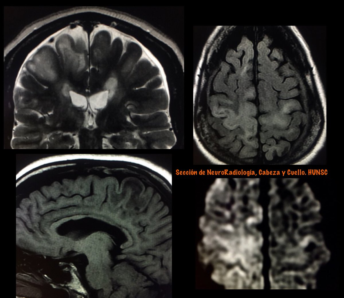
Parasagital meningioma with vasogenic edema
From left to right: coronal T2, enhanced T1 and DWI.
Left side :
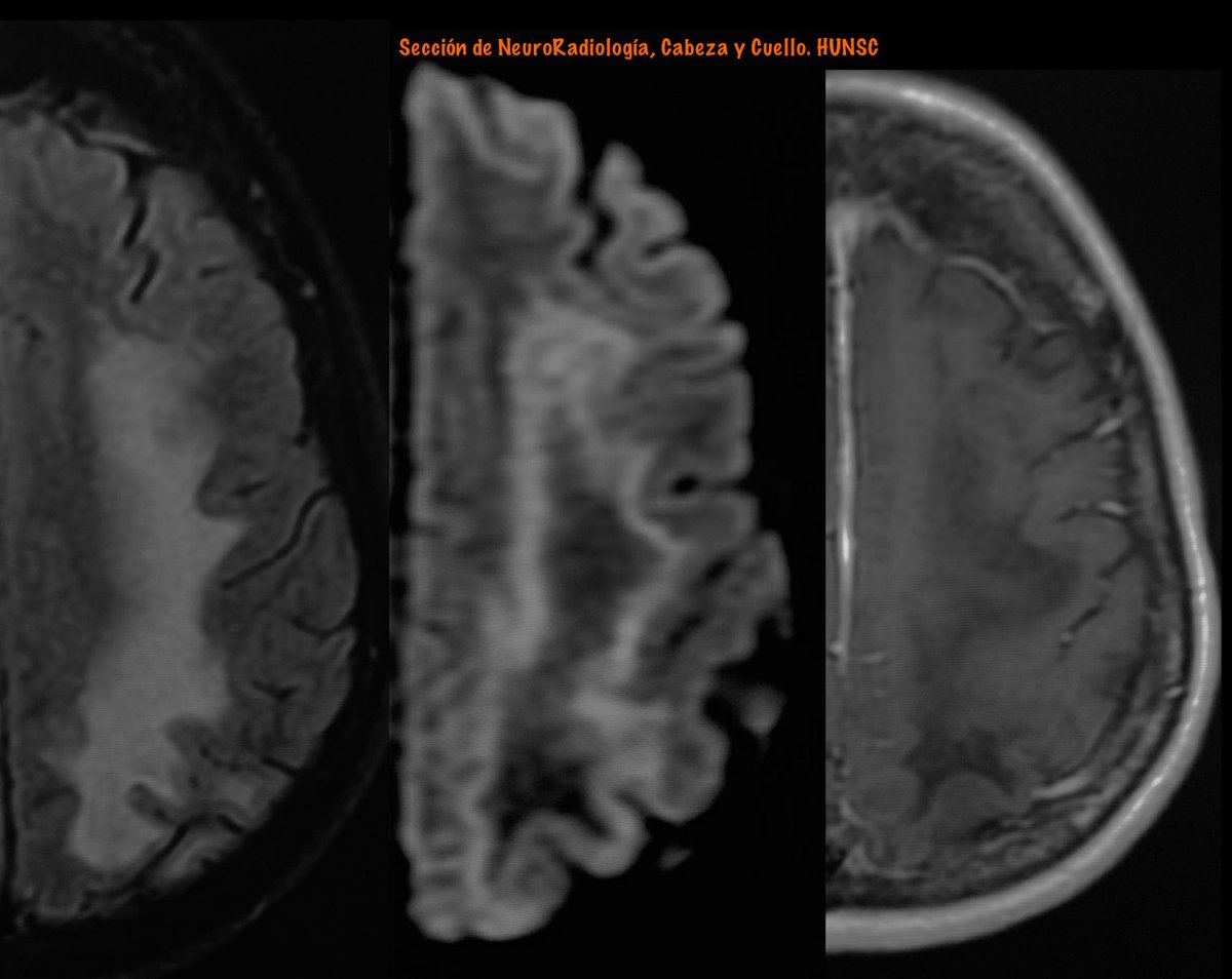
Progressive Multifocal Leukoencephalopathy (PML)
From Left: FLAIR, DWI and enhanced T1 MRI
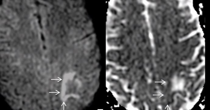
Diffusion Imaging In PML:
The appearance on DWI varies according to the disease stage.
In new active lesions, there is a rim of diffusion restriction at the advancing edge and a central core of facIlitated diffusion.
The rim is usually incomplete and signifies active infection.
Histopathologically, this advancing edge correlates with large swollen oligodendrocytes, enlarged “bizarre astrocytes” with numerous large processes, and infiltration of foamy macrophages. This cellular enlargement constricts the extracellular space.
AJNR 2010
DOI 10.3174/ajnr.A2035
10.1594/ecr2015/B-0300
Neurology 2016;86;1516-1523
DOI 10.1212/WNL.0000000000002586
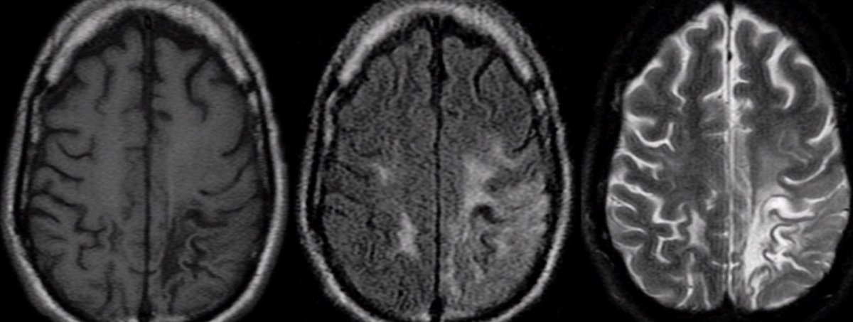
Primary Progressive Multiple sclerosis (PPMS).
A MS course characterised by steadily increasing from disease onset.
Objectively documented neurological disability independent of relapses.
Fluctuations, periods of stability, and superimposed relapses might occur
Primary Progressive Multiple Sclerosis can be diagnosed in patients with:
• 1 year of disability progression (retrospectively or prospectively determined) independent of clinical relapse
Plus two of the following criteria
• One or more T2-lesions characteristic of MS in one or more of the following brain regions: periventricular, cortical or juxtacortical, or infratentorial
• Two or more lesions in the spinal cord
• Presence of CSF-oligoclonal bands
Infarction of the Choroid Plexus
This unusual pattern of ischemia likely resulted from involvement of a medial posterior choroidal artery, emanating from the posterior cerebral artery in close proximity to the thalamogeniculate perforators that caused the thalamocapsular lacune.
Usually have no clinical significance.
Key Diagnostic Features:
Unilateral diffusion restriction of the choroid plexus with evolution with time as expected for infarction.
The association of another infarction in the same vascular territory helps in the diagnosis.
Differential Diagnoses:
Choroid plexus xanthogranuloma:
Variable signal characteristics depending on the mixture of lipid, fluid, and blood products; does not fully suppress on FLAIR; high signal on DWI, usually bilateral.
#FOAMrad#neurorad#HUNSC#daybydaycases
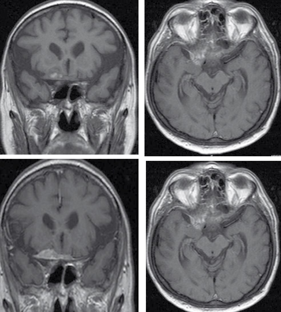
Two patients with a similar clinical profile: elderly men with prostatic adenocarcinoma.
Similar neuroimaging pattern: dural base lesion with intense enhancement and vasogenic edema.
But with different etiology .....
A: Meningioma
B: Dural metastases (DM)
DM in isolation is rare, comprising <1% of all intracranial metastases, and most commonly originates from renal, lung, and breast cancer, as well as carcinoid, adenoid cystic carcinoma, prostatic adenocarcinoma (PCa), and dermatofibrosarcoma
Transdural metastases may mimick meningiomas in radiologic imaging, especially in patients with undiagnosed symptoms of prostatism or PCa.
In older men with dural lesion(s), a possibility of PCa metastases must be considered and radiologic evaluation
#FOAMrad#radres#neurorad
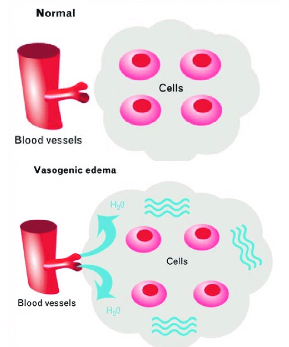
Cerebral Edema
Is an excess accumulation of water in the intracellular and/or extracellular spaces of the brain
Is a common response to various forms of brain injury, and the causes can be categorized as cytotoxic, vasogenic, interstitial, or combined
Cytotoxic edema
• Is cell swelling caused by damaged molecular sodium–potassium ATPase ion pumps. It can affect both gray and white matter.
• Is caused by cell death, most commonly due to infarct or excitotoxic (secondary to excessive neurotransmitter stimulation) brain injury.
Vasogenic edema
• Is interstitial edema caused by increased capillary permeability, by breakdown of the tight endothelial junctions comprising the BBB, secondary to either physical disruption or release of vasoactive compounds
• It is seen primarily in the white matter
PML in MS
Progressive multifocal leukoencephalopathy (PML) is an opportunistic infection of the CNS caused by John Cunningham virus (JCV)
PML in the setting of monoclonal antibody therapy has attracted tremendous attention particularly in MS patients treated with natalizumab/NTZ.
Features favoring PML over MS (as proposed by Yousry et al) are the following: 1) Diffuse subcortical rather than periventricular white matter involvement; frequent involvement of posterior fossa. 2) Irregular ill-defined infiltrating edge confined to the white matter.
3) Persistent progression of the lesion confined within the white matter tract. 4) No mass effect even in large lesions. 5) Diffuse increased T2 signal intensity; recently involved areas more T2 hyperintense than the old areas.