Primary Progressive Multiple sclerosis (PPMS).
A MS course characterised by steadily increasing from disease onset.
Objectively documented neurological disability independent of relapses.
Fluctuations, periods of stability, and superimposed relapses might occur
Primary Progressive Multiple Sclerosis can be diagnosed in patients with:
• 1 year of disability progression (retrospectively or prospectively determined) independent of clinical relapse
May 3, 2021 • 6 tweets • 4 min read
#FOAMrad#neurorad#radres#radmed#HUNSC#daybydaycases#radiology#MedEd
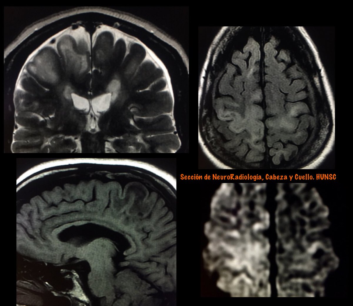
One patient/two sides……and two pathologies.
From left to right axial FLAIR, coronal T2 and axial T1 MRI
Right side:
Parasagital meningioma with vasogenic edema
From left to right: coronal T2, enhanced T1 and DWI.
Jun 8, 2020 • 5 tweets • 3 min read
Infarction of the Choroid Plexus
This unusual pattern of ischemia likely resulted from involvement of a medial posterior choroidal artery, emanating from the posterior cerebral artery in close proximity to the thalamogeniculate perforators that caused the thalamocapsular lacune.
Usually have no clinical significance.
Key Diagnostic Features:
Unilateral diffusion restriction of the choroid plexus with evolution with time as expected for infarction.
The association of another infarction in the same vascular territory helps in the diagnosis.
Feb 3, 2020 • 4 tweets • 3 min read
#FOAMrad#neurorad#HUNSC#daybydaycases
Two patients with a similar clinical profile: elderly men with prostatic adenocarcinoma.
Similar neuroimaging pattern: dural base lesion with intense enhancement and vasogenic edema.
But with different etiology .....
A: Meningioma
B: Dural metastases (DM)
DM in isolation is rare, comprising <1% of all intracranial metastases, and most commonly originates from renal, lung, and breast cancer, as well as carcinoid, adenoid cystic carcinoma, prostatic adenocarcinoma (PCa), and dermatofibrosarcoma
Aug 12, 2019 • 5 tweets • 4 min read
#FOAMrad#radres#neurorad
Cerebral Edema
Is an excess accumulation of water in the intracellular and/or extracellular spaces of the brain
Is a common response to various forms of brain injury, and the causes can be categorized as cytotoxic, vasogenic, interstitial, or combined
Cytotoxic edema
• Is cell swelling caused by damaged molecular sodium–potassium ATPase ion pumps. It can affect both gray and white matter.
• Is caused by cell death, most commonly due to infarct or excitotoxic (secondary to excessive neurotransmitter stimulation) brain injury.
Jun 14, 2019 • 8 tweets • 4 min read
PML in MS
Progressive multifocal leukoencephalopathy (PML) is an opportunistic infection of the CNS caused by John Cunningham virus (JCV)
PML in the setting of monoclonal antibody therapy has attracted tremendous attention particularly in MS patients treated with natalizumab/NTZ.
Features favoring PML over MS (as proposed by Yousry et al) are the following: 1) Diffuse subcortical rather than periventricular white matter involvement; frequent involvement of posterior fossa. 2) Irregular ill-defined infiltrating edge confined to the white matter.